Studying biology or histology can be incredibly frustrating when you hit the chapter on muscle tissues. You mix up smooth, skeletal, and cardiac cells, and suddenly your lab reports or exam grades take a hit. We completely get it. It is easy to get lost in a sea of microscopic terms. We’re here to fix that confusion by breaking down cardiac muscle tissue and heart histology into simple, highly detailed pieces.
Key Takeaways
- Cardiac muscle tissue is an involuntary, striated muscle found exclusively in the walls of the heart.
- The defining features of this tissue are intercalated discs, which physically and electrically connect the cells.
- Cardiomyocytes are unique because they are short, branched fibers that usually contain a single, centrally located nucleus.
Table of Contents
- The Anatomy of the Heart Wall: Where Cardiac Tissue Lives
- Cellular Structure: Inside the Cardiomyocyte
- The Magic of Intercalated Discs
- Mechanism of Synchronized Contractions
- Energy and Endurance: Why the Heart Never Tires
- Cardiac vs. Skeletal vs. Smooth Muscle
- Zoological Heart Morphology and Animal Cardiac Function
- How to Identify Cardiac Tissue Under a Microscope
- Common Histological Anomalies and Diseases
- Frequently Asked Questions
- Wrapping Up Your Histology Journey
The Anatomy of the Heart Wall: Where Cardiac Tissue Lives
Before we look at the microscopic cells, we need to zoom out and look at the heart wall itself. The heart is not just a simple hollow organ. It is a complex, multi-layered pump designed for relentless action. The wall of the heart consists of three distinct layers.
The outermost layer is the epicardium. It provides a protective outer covering and contains connective tissue and fat. It acts as a lubricating layer against the pericardial sac. Then, we have the endocardium. This is the smooth inner lining of the heart chambers. It ensures that blood flows without friction or clotting.
However, the middle layer is the star of the show. We call this the myocardium. The myocardium is entirely made up of cardiac muscle tissue. This is the thickest layer of the heart wall, especially in the left ventricle. The left ventricle has to pump blood to the entire body, so its myocardium is incredibly robust.
According to a 2024 histological research report, over 95% of the heart’s total mass is attributed directly to the thick myocardial layer in healthy adult mammals.
When we talk about heart histology, we are almost entirely talking about the myocardium. This layer is responsible for the pumping action of the heart. Without the thick, muscular myocardium, blood circulation would instantly fail. It is a highly specialized tissue that cannot be found anywhere else in the human body.
💡 Pro Tip: If you are ever asked on an exam where cardiac muscle tissue is located, do not just say ‘the heart’. Specify that it is found in the ‘myocardium of the heart wall’ to secure full marks for accuracy.
Cellular Structure: Inside the Cardiomyocyte
Let’s take a closer look at the individual cells that make up this mighty tissue. We call cardiac muscle cells ‘cardiomyocytes’. Unlike the long, cylindrical fibers of skeletal muscle, cardiomyocytes have a very distinct shape. They are relatively short, measuring about 50 to 100 micrometers in length.
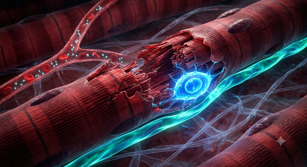
The Branched Appearance
The most noticeable feature of a cardiomyocyte is its branched structure. These cells do not lie perfectly parallel to each other. Instead, they split and branch out, connecting with multiple neighboring cells. This creates a complex, three-dimensional network.
This branching is not just for show. It serves a vital mechanical purpose. By weaving together into a mesh, the cells create a tightly knit fabric. When the heart contracts, this mesh wrings blood out of the chambers efficiently. It is similar to twisting a wet towel to squeeze out the water.
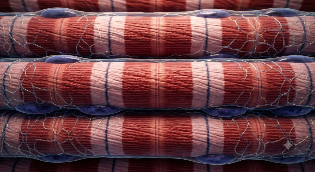
Nucleus and Striations
If you look closely at a cardiomyocyte, you will usually see a single, oval-shaped nucleus. This nucleus sits right in the center of the cell. Occasionally, you might spot a cell with two nuclei, but a single central nucleus is the standard rule. This is a great way to tell it apart from skeletal muscle, which has multiple nuclei pushed to the edges of the cell.
You will also notice faint transverse stripes across the cells. We call these striations. They are caused by the precise arrangement of contractile proteins inside the cell. Specifically, these are actin and myosin filaments organized into repeating units called sarcomeres.
These sarcomeres slide past each other to create a contraction. Because cardiac muscle is striated, it contracts with a lot of force. It shares this powerful sliding filament mechanism with skeletal muscle, but operates entirely on its own schedule.
The Magic of Intercalated Discs
If there is one thing you must remember about heart histology, it is the intercalated discs. These are jagged, dark-staining lines where the ends of two cardiomyocytes meet. They look a bit like a tiny staircase or a zigzag line under a light microscope.
Intercalated discs are essentially super-charged cell junctions. They are the glue that holds the entire myocardium together. Without them, the heart would literally rip itself apart during a vigorous heartbeat. Let’s break down the two main components of an intercalated disc.
Desmosomes: The Physical Anchors
The first major component of the intercalated disc is the desmosome. You can think of desmosomes as microscopic rivets or spot welds. They physically bind the adjacent cells together. When one cell contracts and pulls, the desmosome ensures the neighboring cell doesn’t detach.
There is also a structure here called the fascia adherens. It works alongside the desmosomes. The fascia adherens anchors the actin filaments of the terminal sarcomeres directly to the cell membrane. This transfers the contractile force directly from one cell to the next.
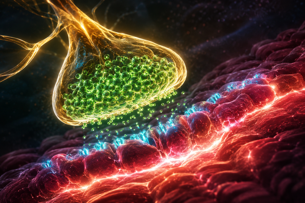
Gap Junctions: The Electrical Highways
While desmosomes provide physical strength, gap junctions provide electrical communication. Gap junctions are tiny protein channels that connect the cytoplasm of one cell directly to the cytoplasm of its neighbor. They allow ions, like sodium and calcium, to flow freely between cells.
This is a massive deal for heart function. When an electrical signal (an action potential) starts in one cell, it instantly shoots through the gap junctions to the next cell. This means the entire network of cells depolarizes and contracts at almost the exact same time.
💡 Pro Tip: A great way to remember this is to think of desmosomes as the physical ‘glue’ and gap junctions as the electrical ‘wires’ connecting the heart cells.
| Intercalated Disc Feature | Primary Function | Analogy |
|---|---|---|
| Desmosomes | Prevents cell separation during contraction. | Spot welds holding metal plates. |
| Fascia Adherens | Anchors actin filaments to the cell membrane. | Ropes tying down a tent. |
| Gap Junctions | Allows rapid ion exchange and electrical signaling. | Open doorways between adjoining rooms. |
Mechanism of Synchronized Contractions
Because of those incredible gap junctions, cardiac muscle tissue behaves as a functional syncytium. This is a fancy biological term meaning that multiple distinct cells act as one single, coordinated unit. When one cell fires, they all fire.
This synchronized contraction is essential for survival. If the heart cells contracted randomly, the heart would just quiver and fail to pump blood. We call that dangerous quivering fibrillation. The functional syncytium ensures a smooth, unified squeeze.
The Role of the Pacemaker
Cardiac muscle is involuntary, meaning you don’t have to consciously think about making it beat. Furthermore, it is highly autorhythmic. The heart generates its own electrical impulses without needing signals from the brain. It does this via specialized cells in the sinoatrial (SA) node.
The SA node acts as the heart’s natural pacemaker. It fires off an action potential, which then ripples through the atrial muscle tissue. The signal then hits the atrioventricular (AV) node, travels down the bundle of His, and spreads through the Purkinje fibers to the ventricles.
Calcium Induced Calcium Release
The actual contraction mechanism inside the cardiomyocyte is fascinating. When the action potential travels down the cell membrane, it dives into deep tubes called T-tubules. This triggers calcium channels to open, letting extracellular calcium into the cell.
This small influx of calcium triggers the sarcoplasmic reticulum inside the cell to release a massive flood of internal calcium. We call this ‘calcium-induced calcium release’. This flood of calcium binds to troponin, moves tropomyosin out of the way, and allows the actin and myosin filaments to slide. The result is a powerful, synchronized heartbeat.
Energy and Endurance: Why the Heart Never Tires
Think about your biceps. If you lift heavy weights, your skeletal muscles eventually get tired and burn with lactic acid. Your heart, on the other hand, beats around 100,000 times a day, every single day of your life. It cannot afford to get tired or take a break.
How does cardiac muscle achieve this tireless, rhythmic nature? The secret lies in its incredible metabolic adaptations. Cardiomyocytes are exclusively designed for aerobic respiration. They demand a constant, heavy supply of oxygen to function.
The Mitochondria Powerhouses
To produce the massive amounts of ATP (energy) required, cardiac muscle cells are packed with mitochondria. In fact, mitochondria take up about 25% to 35% of the total volume of a cardiomyocyte. Compare this to skeletal muscle cells, which might only have 2% mitochondrial volume.
Recent morphological studies in 2023 show that mammalian cardiac tissue contains up to 35% mitochondrial volume, vastly outperforming skeletal muscle endurance and preventing fatigue.
Because of this high reliance on aerobic metabolism, cardiac tissue is extremely rich in myoglobin. Myoglobin is an oxygen-binding protein that stores extra oxygen right inside the cell. It gives the heart muscle its deep, dark red color.
A Rich Capillary Network
To support those mitochondria, the myocardium requires a massive blood supply. The background of any good histological slide of heart tissue will hint at a rich capillary network. These tiny blood vessels weave intimately between the branched muscle fibers.
The coronary arteries constantly feed these capillaries with fresh, oxygenated blood. If this blood flow is ever blocked, the cardiac muscle cells quickly run out of oxygen. Because they cannot switch to anaerobic metabolism efficiently, the cells will begin to die within minutes. This is exactly what happens during a myocardial infarction, or a heart attack.
Cardiac vs. Skeletal vs. Smooth Muscle
One of the most common questions on any biology or medical exam involves comparing the three types of muscle tissue. It is incredibly important to keep their features separated in your mind. Let’s break down the key differences directly.
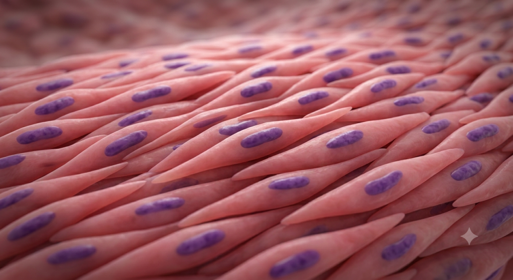
Skeletal muscle is voluntary and striated. It forms long, unbranched cylinders with many nuclei pushed to the edges. Smooth muscle is involuntary and non-striated. It consists of spindle-shaped cells with a single nucleus, found in the walls of your intestines and blood vessels.
Cardiac muscle sits right in the middle. It shares the striations of skeletal muscle but shares the involuntary, single-nucleus nature of smooth muscle. Plus, it has its own unique features like branching and intercalated discs. Let’s look at a clear comparison table.
| Feature | Cardiac Muscle | Skeletal Muscle | Smooth Muscle |
|---|---|---|---|
| Control | Involuntary | Voluntary | Involuntary |
| Striations | Yes | Yes | No |
| Cell Shape | Short, branched | Long, cylindrical | Spindle-shaped |
| Nucleus | Single, central | Multiple, peripheral | Single, central |
| Unique Structures | Intercalated Discs | None | Dense bodies |
💡 Pro Tip: If you are looking at a slide and see striations, you can immediately rule out smooth muscle. From there, look for branching and central nuclei. If you see them, you are definitely looking at cardiac tissue.
Zoological Heart Morphology and Animal Cardiac Function
We often focus solely on the human heart, but comparative zoological heart morphology offers incredible insights. The fundamental structure of cardiac muscle tissue is remarkably conserved across different animal species, but the macro-organization changes drastically.
From Fish to Mammals
In fish, the heart is a simple two-chambered tube. The cardiac muscle pumps blood directly to the gills for oxygenation. Because they live in water and have lower metabolic demands, their myocardium doesn’t need to be as thick as a mammal’s. However, the cells still rely on intercalated discs to function.
Amphibians, like frogs, have a three-chambered heart. Their single ventricle contains highly trabeculated cardiac muscle. These spongy, muscular ridges help separate oxygenated and deoxygenated blood within a single chamber. The cardiomyocytes here are highly adapted to dealing with mixed blood oxygen levels.
The Avian Advantage
Birds have some of the most impressive cardiac muscle tissue in the animal kingdom. Flying requires an unbelievable amount of energy and oxygen. Consequently, bird hearts are proportionally larger and pump much faster than mammalian hearts.
Data from a 2024 comparative zoology index reveals that avian myocardium structure features the highest density of gap junctions among all vertebrates, allowing for ultra-rapid heart rates during flight.
The basic histology remains the same. You will still find branched fibers and a single central nucleus in an avian heart. However, the efficiency of their gap junctions and the density of their capillary networks are scaled up significantly to meet the intense demands of flight.
How to Identify Cardiac Tissue Under a Microscope
Let’s pretend you are sitting in a histology lab. Your professor hands you a glass slide and asks you to identify the tissue. How do you confidently determine that it is a slice of myocardium? There is a specific visual checklist you should follow.
First, scan the tissue at a lower magnification. Look for a general fibrous appearance. You should see a lot of pink or red tissue, assuming standard Hematoxylin and Eosin (H&E) staining has been used. The space between the fibers will likely contain small, flat cells. Those are the endothelial cells of the capillaries.
Spotting the Discs
Next, move to a higher magnification, like 400x. Focus on the individual fibers. You are looking for branching. Do the fibers split like a Y-shape? If yes, you are on the right track. Now, look at the nuclei. Are they sitting right in the middle of the cell cytoplasm? That is another strong indicator.
Finally, search for the ultimate giveaway: the intercalated discs. They will look like thin, dark, transverse bands running perpendicular to the muscle fibers. They are often slightly thicker and darker than the regular striations. If you see those dark, jagged lines, you can confidently declare that you are looking at cardiac muscle tissue.
💡 Pro Tip: Sometimes, H&E staining doesn’t show intercalated discs perfectly. If your lab uses a special stain like Masson’s trichrome or an iron hematoxylin stain, the discs and striations will pop out with much greater contrast.
Common Histological Anomalies and Diseases
Understanding healthy tissue is great, but we also need to understand what happens when things go wrong. Cardiac histology changes significantly under stress or disease conditions. Pathologists look for these microscopic changes to diagnose heart conditions.
Hypertrophy: The Enlarged Heart
When the heart has to work harder than normal—perhaps due to high blood pressure or a faulty valve—the muscle tissue adapts. Unlike skeletal muscle, cardiac muscle cells generally do not divide and multiply to create new cells. We call a lack of cell division ‘amitotic’ behavior.
Instead, the existing cardiomyocytes get larger. They pack in more sarcomeres and grow in diameter. This is called cellular hypertrophy. Under a microscope, hypertrophic cardiac tissue will feature massively thickened, boxcar-like nuclei and unusually wide muscle fibers. While it helps the heart pump harder in the short term, it eventually leads to heart failure.
Ischemia and Scar Tissue
As we mentioned earlier, a lack of blood flow causes cardiac cells to die. When cardiomyocytes die off, the body cannot grow new ones to replace them. Instead, the dead area is invaded by fibroblasts. These are cells that lay down thick, tough collagen fibers to patch the hole.
This process creates a myocardial scar. Under a microscope, you will see a patch of dense, wavy connective tissue completely devoid of muscle fibers. Because scar tissue lacks gap junctions, it cannot conduct electrical signals. This interrupts the functional syncytium and frequently leads to dangerous heart arrhythmias.
Frequently Asked Questions
What is the main function of cardiac muscle tissue?
The primary function is to pump blood continuously throughout the body. It relies on coordinated, involuntary contractions to maintain blood pressure and ensure oxygen reaches all your vital organs without ever stopping to rest.
Why does cardiac muscle have striations?
Striations appear because the cell’s contractile proteins, actin and myosin, are organized into highly structured, repeating patterns called sarcomeres. This organized overlap creates the light and dark bands visible under a microscope and allows for powerful contractions.
Can cardiac muscle tissue repair itself?
No, it has an extremely limited capacity for repair. Once adult cardiomyocytes die from injury or a heart attack, they do not regenerate. The body replaces the dead tissue with non-contractile, fibrous scar tissue instead.
What happens if gap junctions fail?
If gap junctions fail, electrical signals cannot pass rapidly between cells. The heart loses its ability to contract as a single unit. This leads to disorganized, ineffective twitching known as fibrillation, which stops blood flow entirely.
How is cardiac muscle controlled if it is involuntary?
While the heart generates its own beat via pacemaker cells, the autonomic nervous system speeds it up or slows it down. Sympathetic nerves increase your heart rate during stress, while parasympathetic nerves slow it down during rest.
Is cardiac muscle found in veins or arteries?
No. Cardiac muscle is found exclusively in the myocardium, the middle layer of the heart wall. Your blood vessels contain smooth muscle tissue, which helps regulate blood pressure by constricting or dilating the vessels.
Wrapping Up Your Histology Journey
You have now explored the intricate world of cardiac muscle tissue. We’ve examined the unique branched cardiomyocytes, uncovered the essential role of intercalated discs, and discussed how mitochondria power this tireless biological engine. You understand exactly how desmosomes and gap junctions work together to create a perfectly synchronized heartbeat. Studying histology doesn’t have to be a nightmare when you break it down into functional concepts rather than just memorizing terms.
It is genuinely fascinating how microscopic structures dictate the survival of the entire organism. From zoological comparisons to human pathology, the myocardium stands out as a uniquely resilient tissue. Now that you are a master of heart histology, we want to hear from you. Which feature of the cardiac muscle cell do you find the most interesting, and why? Let us know in the comments section below!