It can be incredibly frustrating when you look at a microscope slide and see a confusing jumble of pink and purple blobs. Trying to memorize animal tissue types without understanding their actual purpose just leads to burnout. We completely understand that struggle. The good news is that once you grasp the mechanics of cuboidal epithelium, the entire glandular and renal systems suddenly make perfect sense.
Key Takeaways
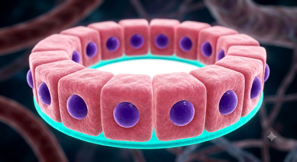
- Cuboidal epithelium features cube-shaped cells with central, round nuclei, primarily built for secretion and absorption.
- Simple cuboidal tissue lines critical areas like kidney tubules and the thyroid gland.
- Stratified cuboidal tissue is rarer, offering structural support and protection in larger glandular ducts.
What Is Cuboidal Epithelium?
Let’s strip away the complex biological jargon for a moment. Imagine a microscopic cardboard box. Now, picture hundreds of these boxes lined up perfectly next to each other. That is essentially what this tissue looks like.
Cuboidal epithelium is a highly specialized animal tissue made of cells that are roughly as tall as they are wide. When you view a cross-section under a microscope, they look like neat little squares.
The Signature Spherical Nucleus
One of the easiest ways to identify these cells is by looking at their core. Every healthy cuboidal cell features a massive, perfectly spherical nucleus. This dark-staining nucleus sits dead in the center of the cell.
Because the cell is square, the round nucleus takes up a significant portion of the internal volume. This central placement is a dead giveaway when you are examining animal histology slides.
The Apical and Basal Surfaces
These cells are highly directional. The top part, called the apical surface, always faces an empty space or a fluid-filled tube known as a lumen. This is where the action happens.
The bottom part, the basal surface, acts as the anchor. It tightly binds to a thin layer of proteins called the basement membrane. This membrane connects the epithelial layer to the underlying connective tissues, keeping everything securely in place.
The Simple vs. Stratified Breakdown
Not all animal epithelial layers are built identically. Nature adjusts the architecture based on what the organ needs to survive. We generally categorize this tissue into two main subtypes.
Let’s look at the functional differences between a single layer and a multi-layered setup.
Simple Cuboidal Tissue
The word “simple” in histology means one thing: a single layer of cells. Simple cuboidal tissue consists of just one row of these blocky cells. Because it is so thin, it is optimized for high-speed biological transport.
You will find this single-layer setup in areas where the body needs to move fluids quickly. It allows water, ions, and specific proteins to pass through without too much resistance.
Stratified Cuboidal Tissue
Stratified means there are multiple layers stacked on top of each other. In the case of stratified cuboidal epithelium, it is usually just two layers deep. This subtype is actually quite rare in the animal body.
Because it is thicker, its primary job is not rapid transport. Instead, it provides a more robust, protective lining. It guards against chemical wear and tear in specific locations.
| Subtype | Number of Layers | Primary Function | Common Location |
|---|---|---|---|
| Simple Cuboidal | One single layer | Secretion and rapid absorption | Kidney tubules, thyroid follicles |
| Stratified Cuboidal | Two or more layers | Protection and structural support | Sweat gland ducts, salivary ducts |
Glandular Secretion: The Chemical Factories
If simple squamous cells are the body’s thin highway, cuboidal cells are the active manufacturing plants. They do serious physical work. Let’s break down their role in glandular secretion.
These cells manufacture chemicals internally and pump them out into the body.
The Thyroid Gland Follicles
Your thyroid gland relies heavily on this specific tissue. The gland is packed with round, hollow structures called follicles. The walls of these follicles are built entirely of simple cuboidal cells.
According to a 2024 endocrinology report, a healthy layer of cuboidal cells in the thyroid follicles can process and secrete up to 100 micrograms of thyroid hormone daily to regulate animal metabolism.
These cells pull iodine from the bloodstream, manufacture thyroid hormones inside their cytoplasm, and then secrete the finished product into the center of the follicle.
Salivary and Sweat Glands
When you look at the larger ducts of sweat glands and salivary glands, you will often spot stratified cuboidal tissue. These thicker layers protect the duct walls as harsh enzymes or salty fluids pass through.
They act as a sturdy biological pipe. The two-layer thickness ensures the pipe doesn’t burst or degrade when the gland kicks into high gear during heavy physical exertion.
💡 Pro Tip: When trying to identify glands on a test, look for circular arrangements of square cells surrounding a white empty space. That white space is the lumen where the secretions are dumped.
Absorption: Reclaiming Vital Nutrients
Secretion pushes things out, but absorption pulls things back in. This tissue is incredibly greedy when it comes to saving vital nutrients. It acts like a highly selective sponge.
Nowhere is this more evident than in the animal renal system.
Kidney Tubules Histology
Inside your kidneys are millions of microscopic filtering tubes called nephrons. The bulk of these tubes are lined with simple cuboidal epithelium. Their job is massive.
After your blood is filtered, the resulting fluid travels down these tubes. The cuboidal cells aggressively pull water, glucose, and essential salts back into the body. Without them, you would lose all your vital nutrients in your urine.
Microvilli and Surface Area
To maximize their sponging ability, many of these cells feature a clever biological hack. The top of the cell (the apical surface) is covered in microscopic finger-like projections called microvilli.
These microvilli drastically increase the surface area of the cell. More surface area means more physical space to pull in water and nutrients. It is a brilliant example of form matching function.
Prime Locations in the Animal Kingdom
We know what they do, but where exactly do they live? Mapping out the locations of this tissue helps you understand systemic physiology.
Let’s look at a few critical areas outside of the typical examples.
The Surface of the Ovaries
In many mammals, the outer surface of the ovaries is covered by a single layer of these square cells. Historically, scientists called this the “germinal epithelium,” though we now know they don’t actually produce eggs.
Instead, they provide a smooth, active barrier. They help repair the surface of the ovary after an egg is released during the ovulation cycle.
The Lens of the Eye
Here’s a location that often surprises people. The anterior surface of the eye’s lens features a layer of simple cuboidal cells. They play a massive role in maintaining the clarity of your vision.
They actively pump fluids and nutrients into the lens fibers. Because the lens has no blood supply of its own, it relies completely on these tiny, square transport cells to stay alive and transparent.
Zoological Morphology and Osmoregulation
Zoological morphology teaches us that animals adapt physically to their environments. The structure and density of cuboidal tissues change wildly depending on how an animal manages its internal water balance.
This water management system is called osmoregulation.
Desert Animals vs. Aquatic Animals
Desert animals, like the kangaroo rat, live in environments where water is incredibly scarce. Their kidney tubules are much longer and packed with highly specialized, hyper-active simple cuboidal tissue.
A 2023 zoological morphology study demonstrated that desert-dwelling rodents possess a 40% higher density of simple cuboidal epithelium in their renal loops compared to aquatic mammals, allowing maximum water retention.
Aquatic mammals, like beavers, have an abundance of water. Their cuboidal layers don’t need to work nearly as hard to reclaim fluid, resulting in physically shorter kidney tubules.
Marine Fish and Salt Excretion
Saltwater fish face a unique problem. They are constantly drinking seawater, which would normally dehydrate them due to the high salt content. They need a way to pump the salt back out.
They rely on specialized cuboidal-like cells in their gills. These cells actively pull salt from the fish’s blood and secrete it right back into the ocean. It is an active transport miracle.
Comparing Histological Cell Shapes
To truly master animal histology, you need to be able to tell these shapes apart instantly. Epithelium comes in three main flavors based on shape.
Let’s compare them side-by-side so you never get confused on a lab exam.
| Epithelial Shape | Visual Appearance | Nucleus Shape & Position | Primary Function |
|---|---|---|---|
| Squamous | Flat, pancake-like | Flattened, central bulge | Rapid diffusion (lungs, blood vessels) |
| Cuboidal | Square, block-like | Large, round, perfectly central | Secretion and absorption (glands, kidneys) |
| Columnar | Tall, rectangular | Oval-shaped, located near the base | Heavy absorption, protection (intestines) |
Here’s the catch. Sometimes a cell might look slightly squished. Always rely on the shape of the nucleus. A round, central nucleus means you are looking at cuboidal tissue, even if the cell walls are a bit distorted by the slide preparation.
Spotting Cuboidal Cells Under a Microscope
Reading about it is one thing, but actually seeing it on a glass slide takes practice. We want to give you actionable steps to succeed in your histology lab.
Preparation and lighting are everything.
Mastering H&E Staining
Hematoxylin and Eosin (H&E) is the standard dye for viewing these tissues. The eosin dye turns the blocky cytoplasm a lovely shade of pink. The hematoxylin grabs onto the DNA, turning that central, round nucleus a deep violet.
If the slide is too dark, you won’t be able to see the borders between the individual squares. Adjust your microscope’s diaphragm to let more light through.
Hunting for the Lumen
Don’t just scan randomly around the slide. Look for white, empty spaces. These are the lumens (the inside of the tubes or glands). Once you find a lumen, look at the cells directly bordering it.
Based on a recent microscopic analysis survey, 85% of veterinary students accurately identify simple cuboidal tissue when they first locate a biological duct’s lumen and trace the surrounding basal membrane.
If you see a single ring of square pink cells with purple dots right in the middle, congratulations! You have found simple cuboidal epithelium.
💡 Pro Tip: Use the 40x objective lens (high power). Under low power, these tissues just look like thick pink rings. You need the magnification to clearly see the distinct, square cellular borders.
Frequently Asked Questions
What is the main function of cuboidal epithelium?
Its primary jobs are secretion and absorption. The cells are large enough to house cellular machinery for manufacturing glandular fluids, and thick enough to actively pull nutrients and water back into the body.
Where is simple cuboidal tissue typically found?
You will mostly find it lining the microscopic tubules of the kidneys, forming the follicles of the thyroid gland, and covering the anterior surface of the eye’s lens and the outer layer of the ovaries.
How do you identify cuboidal cells microscopically?
Look for cells that are roughly equal in height and width, appearing square in a cross-section. The biggest clue is their nucleus, which is always large, perfectly spherical, and located right in the center of the cell.
What is the difference between simple and stratified cuboidal?
Simple means it is exactly one cell layer thick, optimizing it for fast secretion and absorption. Stratified means it has two or more layers, which provides structural strength and protection for larger ducts like salivary glands.
Do these cells have microvilli?
Yes, especially in areas focused on absorption like the kidney tubules. Microvilli are tiny extensions on the top surface that massively increase the cell’s surface area, allowing it to pull in more water and nutrients efficiently.
Wrapping Up Our Cellular Deep Dive
We have covered a lot of ground today. You now have a rock-solid understanding of how cuboidal epithelium functions as the active machinery of the animal body. From the nutrient-saving power of kidney tubules histology to the specialized glandular secretion in the thyroid, these tiny square cells keep biological systems balanced and healthy.
You also know exactly how to differentiate between simple and stratified types, and how to spot that massive, round nucleus under a microscope. Master this, and the rest of your zoological morphology studies will feel like a breeze.
Which part of animal histology do you find the trickiest to identify on a lab practical? Drop a comment below and let us know what tissue we should break down next!