Studying histology can feel completely overwhelming. You stare at microscope slides, struggling to tell one pink cellular blob from another, fearing you’ll fail your upcoming anatomy exam. We have all been there. By mastering columnar epithelium, you’ll finally understand how our bodies absorb vital nutrients and physically fight off infections.
Key Takeaways
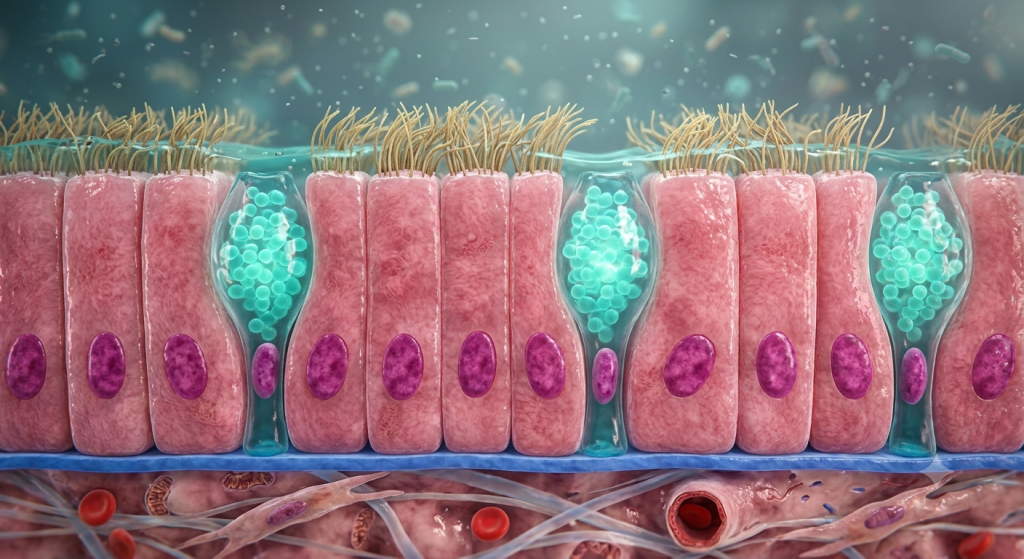
- Columnar epithelium consists of tall, rectangular cells with distinct oval nuclei located near the basal membrane.
- Non-ciliated forms utilize microvilli to maximize nutrient absorption, primarily lining the digestive tract.
- Ciliated forms act as tiny biological brooms, sweeping mucus and debris through the respiratory tract and reproductive systems.
What Is Columnar Epithelium?
Let’s break down the basic architecture first. Columnar epithelium gets its name from its distinct physical shape. These cells look like tall, sturdy columns or rectangles standing shoulder-to-shoulder. They are significantly taller than they are wide.
When you look closely at animal tissue structures, you notice something specific about their nuclei. Instead of being perfectly round and centered, the nuclei in columnar cells are elongated ovals. Furthermore, they are almost always located near the bottom of the cell.
The Basal and Apical Surfaces
Every epithelial cell has a top and a bottom. The bottom is called the basal surface. It firmly attaches to the underlying basement membrane, anchoring the tissue to the rest of the body. The top is the cellular apical surface.
The apical surface faces the open space, or lumen, of the organ it lines. This is where all the heavy lifting happens. Depending on the organ’s needs, nature modifies this top surface with highly specialized structures.
Non-Ciliated Columnar Epithelium: The Absorptive Powerhouse
Let’s talk about the digestive system. Here, the primary goal isn’t just moving things around; it is aggressive absorption. The non-ciliated columnar epithelium is perfectly engineered for this exact job.
You’ll find this specific tissue lining most of your digestive tract, running from your stomach all the way down through your intestines. It acts as a highly selective biological sponge.
The Magic of Microvilli
While this tissue lacks cilia, it is far from bare. The cellular apical surface is densely packed with microvilli. These are microscopic, finger-like projections of the cell membrane itself. They don’t move independently.
According to a 2024 histological database report, a single absorptive columnar cell in the human small intestine can possess up to 3,000 microvilli, expanding its surface area by over 20 times.
By drastically increasing the surface area, microvilli allow the cell to suck up massive amounts of nutrients, water, and electrolytes from your digested food. Without them, we would quickly starve regardless of how much we ate.
💡 Pro Tip: On a microscope slide, a dense layer of microvilli looks like a fuzzy border at the top of the cells. Histologists commonly refer to this as a “brush border.”
Ciliated Columnar Epithelium: The Movement Masters
Now, let’s switch gears and look at environments where physical movement is necessary. Ciliated columnar cells have a totally different top-level design. Instead of stubby microvilli, they grow long, hair-like structures called cilia.
These cilia are highly active. They beat in coordinated, wave-like rhythms. Think of a massive crowd doing “the wave” in a sports stadium. That is exactly how cilia function.
Protecting the Respiratory Tract Tissue
Your respiratory tract tissue relies heavily on this ciliated setup. When you breathe in dust, bacteria, or pollutants, they get trapped in a sticky layer of mucus. The ciliated columnar cells then go to work.
They constantly sweep this contaminated mucus upward, away from your delicate lungs and toward your throat. Once it reaches the top, you either cough it out or swallow it down into the acidic stomach. It is your body’s built-in escalator for trash removal.
Movement in the Reproductive System
We also find ciliated columnar epithelium in the female reproductive system, specifically lining the fallopian tubes. Here, the cilia have a very delicate job.
After an ovary releases an egg, it cannot move on its own. The gentle, rhythmic beating of the cilia creates a physical fluid current. This current literally sweeps the egg down the tube toward the uterus for potential fertilization.
The Unsung Heroes: Goblet Cells and Mucus Production
You cannot discuss columnar epithelium without mentioning its best friend: the goblet cell. These specialized cells are scattered right alongside the tall columnar cells. They look like tiny wine goblets, which is how they got their name.
Their entire existence revolves around one task: manufacturing and releasing mucus.
The Chemistry of Mucus
Goblet cells produce mucin, a complex glycoprotein. When mucin mixes with water outside the cell, it rapidly expands and transforms into the thick, sticky substance we know as mucus.
Based on a 2023 gastroenterology tissue study, the goblet cells in a healthy human colon secrete roughly 1 to 1.5 liters of protective mucus every single day.
This goblet cells mucus provides essential lubrication. In the digestive tract histology, it allows solid waste to slide through smoothly. In the respiratory system, it forms the sticky trap that catches airborne invaders.
Comparing the Apical Surfaces: Microvilli vs. Cilia
It is easy to get microvilli and cilia confused. Both sit on top of the cells, and both are microscopic. However, their internal structures and core functions are completely different.
Let’s look at the hard data to clear up any confusion.
| Feature | Microvilli (Non-Ciliated) | Cilia (Ciliated) |
|---|---|---|
| Structure | Short, tightly packed folds of the cell membrane. | Long, hair-like projections containing microtubules. |
| Primary Function | Increases surface area for maximum absorption. | Creates movement to sweep fluids and particles. |
| Active Movement? | No. They are entirely stationary. | Yes. They beat actively using cellular energy (ATP). |
| Primary Location | Small intestine, stomach, gallbladder. | Trachea, bronchi, fallopian tubes. |
Here’s the catch. You will never find large amounts of cilia where intense nutrient absorption is needed. The energetic cost of beating cilia would waste the energy the cell is trying to absorb.
Histology Under the Microscope: Spotting the Differences
So, you are sitting in a lab staring down the barrel of a microscope. How do you actually identify these tissues in real life? It takes a bit of practice and an understanding of tissue staining.
Most slides use Hematoxylin and Eosin (H&E) stains. This turns the cell bodies pink and the nuclei a dark, bold purple.
Finding the Basal Nuclei
First, find the edge of the tissue facing the empty white space (the lumen). Look at the shape of the cells forming that border. Are they tall and skinny? Good.
Next, check the purple nuclei. If they are stretched out into ovals and lined up neatly near the bottom half of the cells, you are definitely looking at columnar epithelium.
Spotting the Goblet Cells
Goblet cells are wonderfully easy to spot. Because standard H&E stains do not attach well to mucus, the goblet cells often look like clear, empty white bubbles wedged between the pink columnar cells.
💡 Pro Tip: If you see tall cells with a blurry “brush border” on top and lots of clear white bubbles scattered throughout, you are almost certainly looking at a slide of the small or large intestine.
Real-World Clinical Significance
Why does any of this matter? Because when these cellular structures fail, severe diseases occur. Your health relies entirely on the structural integrity of your epithelial layers.
Let’s look at what happens when things go wrong in both ciliated and non-ciliated tissues.
Celiac Disease and Microvilli Destruction
Celiac disease is a severe autoimmune condition triggered by eating gluten. When a person with Celiac eats wheat, their immune system mistakenly attacks the non-ciliated columnar epithelium lining their small intestine.
This attack literally destroys the microvilli, flattening the cells out. Without that extra surface area, the patient cannot absorb nutrients, leading to severe malnutrition, bloating, and fatigue.
Cystic Fibrosis and the Mucus Escalator
In the respiratory tract, ciliated cells need the mucus to be thin and watery to sweep it away. Cystic fibrosis is a genetic disease that causes goblet cells to produce incredibly thick, dehydrated mucus.
A recent 2025 respiratory health index indicates that over 70% of recurrent lung infections in cystic fibrosis patients stem directly from the failure of ciliated columnar cells to clear ultra-dense mucus.
The cilia simply aren’t strong enough to move this concrete-like mucus. Bacteria get trapped and multiply, leading to chronic, life-threatening lung infections.
Frequently Asked Questions
What is the main function of columnar epithelium?
Its primary functions include intense absorption of nutrients and the secretion of protective mucus. Depending on whether it has cilia, it also acts as a physical transport system for moving fluids, eggs, or debris across its surface.
Where do you find ciliated columnar epithelium?
You will mostly find this specialized ciliated tissue lining the upper respiratory tract (like the trachea and bronchi) and the female reproductive system (specifically the fallopian tubes).
What exactly do goblet cells do?
Goblet cells act as tiny, single-celled glands. They continuously synthesize and secrete mucin, which mixes with water to form the sticky mucus that lubricates the digestive tract and protects the respiratory system.
How do you tell the difference between cilia and microvilli?
Microvilli are short, stationary folds of the cell membrane used strictly for absorbing nutrients. Cilia are much longer, actively moving hair-like structures used to physically sweep materials across the top of the tissue.
Why are the nuclei at the bottom of columnar cells?
By keeping the bulky nucleus near the basal membrane, the cell frees up space in its upper regions. This allows more room for cellular machinery dedicated to secretion (in goblet cells) or absorption processing.
Wrapping Up Our Tissue Tour
We have covered massive ground today regarding animal tissue structures. You now understand that columnar epithelium isn’t just a static wall of cells. Whether it is the non-ciliated cells absorbing your breakfast or the ciliated forms keeping your lungs clear, this tissue is incredibly dynamic.
You also know exactly how to identify these structures, including the vital goblet cells mucus factories, under a microscope. This knowledge bridges the gap between basic cell biology and complex human health.
Which type of epithelial tissue do you find the most fascinating to study under a microscope? Let us know your thoughts in the comments below!