Studying animal histology can be incredibly frustrating when you cannot visualize how microscopic structures drive massive biological processes. You stare at textbook diagrams, but the sheer complexity of cellular diffusion and tissue layers just leaves you confused. We get it. The good news is that mastering the simple squamous epithelium makes the rest of anatomy click into place perfectly. Once you understand this foundational tissue, everything from breathing to kidney function makes complete sense.
Key Takeaways
- Simple squamous epithelium consists of a single layer of flat, scale-like cells designed for rapid transport.
- Its primary functions include cellular diffusion, passive filtration, and fluid secretion in serous membranes.
- Zoological morphology shows this tissue adapts uniquely across species, optimizing oxygen exchange in both mammals and amphibians.
What Exactly Is Simple Squamous Epithelium?
Let’s talk about the absolute foundation of biological tissues. The simple squamous epithelium is exactly what its name suggests. It is a single, incredibly flat layer of cells. Think of it like a tiled floor in a laboratory.
Each cell fits tightly against its neighbor, leaving almost no gap between them. This creates a smooth, continuous, and highly permeable surface. It is the minimalist architecture of the biological world.
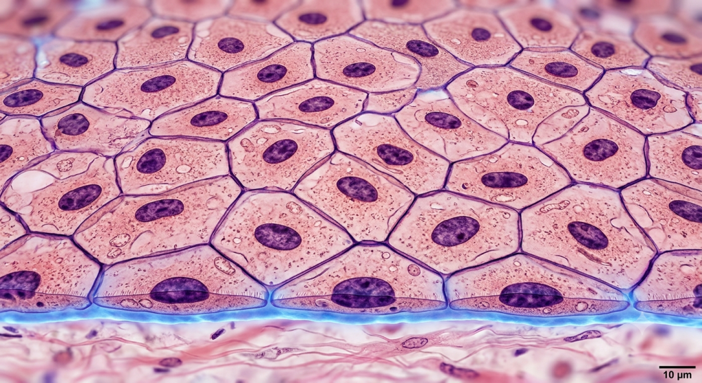
A Microscopic Look at the Squamous Cells
When you look at these squamous cells under a powerful microscope, they look remarkably like fried eggs. The main body of the cell, the cytoplasm, is incredibly thin and spread out. Right in the middle, you will find a flattened, dark-staining nucleus.
This nucleus bulges slightly upwards. Because the cell itself is so thin, the nucleus is often the only visible feature under low magnification. You have to adjust your microscope’s fine focus just to see the edges of the cell membrane.
The Role of the Basement Membrane
Every epithelial tissue needs a foundation. For this specific tissue type, that foundation is called the basement membrane. It is a thin, non-cellular layer made of protein fibers and glycoproteins.
This membrane glues the fragile epithelial cells to the underlying connective tissue. Without this anchor, the single layer of cells would simply tear apart under the slightest physical stress. It also acts as a selective biological filter.
The Core Functions: How It Keeps Animals Alive
Nature never designs something without a specific purpose. Because this tissue is so incredibly thin, it does not offer much protection against physical trauma. Instead, it prioritizes speed and efficiency.
Its thinness is its greatest asset. By minimizing the distance between two environments, it allows materials to pass through rapidly. Let’s break down the exact mechanisms.
Rapid Cellular Diffusion Explained
Cellular diffusion is the movement of molecules from an area of high concentration to low concentration. This tissue is the undisputed champion of this process. Gases like oxygen and carbon dioxide pass right through the cell membranes.
According to a 2024 histological imaging report, oxygen diffusion across the simple squamous epithelium in human alveoli occurs in less than 0.25 seconds under resting conditions.
This rapid exchange keeps our blood oxygenated. If these cells were any thicker, you would literally suffocate, no matter how hard you breathed.
Filtration Mechanisms in the Body
Besides gases, this tissue is exceptional at filtering fluids. In the kidneys, blood pushes against these thin cells under high pressure. Water and small solutes squeeze through the microscopic gaps.
Large proteins and blood cells stay trapped inside the blood vessels. This passive filtration is the very first step in making urine and clearing toxins from the animal body.
Secretion in Serous Membranes
Diffusion and filtration are passive, but these cells also do active work. They secrete a slippery, lubricating fluid. We find this in the serous membranes that line our internal body cavities.
This fluid prevents friction. When your heart beats or your lungs expand, they slide smoothly against the surrounding tissues. Without this secretion, our organs would literally rub themselves raw.
Key Locations in the Animal Body
You will only find simple squamous epithelium where rapid transport or friction reduction is absolutely necessary. It is never found in areas exposed to harsh wear and tear, like the skin.
Let’s map out exactly where this tissue lives inside the body. Knowing these locations helps you understand systemic physiology.
The Alveoli of the Lungs
The lungs are packed with millions of tiny air sacs called alveoli. The walls of these sacs are made entirely of simple squamous cells. This is where the magic of respiration happens.
Oxygen fills the sac and instantly diffuses across the thin cellular barrier into the blood. At the exact same time, carbon dioxide diffuses out. It is a brilliant, high-speed exchange system.
Kidney Glomeruli and Bowman’s Capsule
Inside your kidneys, there are millions of microscopic filtration units called nephrons. At the start of each nephron is a bundle of capillaries surrounded by a structure called Bowman’s capsule.
The outer wall of this capsule is composed of our flat squamous cells. They form a perfect, leaky cup that catches the filtered blood plasma before sending it down the kidney tubules.
Blood Vessel Linings (Endothelium)
Here is a fascinating fact. The entire cardiovascular system is lined with this tissue. From the inside of your heart to the tiniest capillary, there is a continuous layer of flat cells.
When it lines blood vessels, we call it the endothelium. It creates an ultra-smooth surface that prevents blood clots. It also controls exactly what nutrients exit the bloodstream to feed the surrounding muscles.
Zoological Morphology: Species Adaptations
Zoological morphology teaches us that tissues adapt to fit the environment. While humans have highly efficient squamous tissues, other animals have evolved fascinating variations. Let’s look at how nature tweaks this design.
Different species face different metabolic demands. A frog’s tissue requirements differ wildly from a soaring eagle’s needs.
Amphibian vs. Mammalian Adaptations
Amphibians like frogs have very simple lungs. However, they can also breathe directly through their skin. Their internal simple squamous linings are highly vascularized to support this dual breathing method.
Mammals, on the other hand, rely entirely on the massive surface area of their alveoli. Our squamous cells are specialized tightly for internal gas exchange, entirely abandoning the skin-breathing route.
Avian Respiratory Efficiency
Birds have the most efficient respiratory systems on the planet. They need massive amounts of oxygen to sustain flight. Their lungs feature specialized air capillaries instead of our round alveoli.
A 2023 veterinary morphology study found that avian respiratory systems feature simple squamous barriers that are up to 30% thinner than mammalian counterparts, maximizing flight endurance.
This ultra-thin barrier allows birds to fly at high altitudes where oxygen levels are incredibly low. It is a biological engineering marvel.
Histology Techniques: Seeing the Unseen
You cannot just put a piece of raw tissue under a microscope and expect to see clear cellular structures. You have to prepare it properly. Let’s walk through how scientists actually study these cells.
It takes precision and the right chemicals to reveal the hidden world of animal histology.
H&E Staining Basics
Hematoxylin and Eosin (H&E) is the gold standard for tissue staining. Hematoxylin is a dark blue dye that binds specifically to the nucleus. Eosin is a pink dye that colors the cytoplasm and extracellular matrix.
When you apply H&E to simple squamous epithelium, the flat, pancake-like cells turn a soft pink. The flattened central nuclei pop out in a bold, dark purple. It makes identifying the single layer incredibly easy.
💡 Pro Tip: If your H&E slide looks completely pink with no blue nuclei, you likely over-washed the slide in alcohol, stripping away the hematoxylin dye. Always time your washes perfectly!
Advanced Electron Microscopy
Standard light microscopes are great for basic viewing, but they have limits. To see the tiny pores and junctions between squamous cells, scientists use Scanning Electron Microscopes (SEM).
SEM blasts the tissue with a beam of electrons. This creates a highly detailed, 3D image of the tissue’s surface. It allows researchers to see exactly how these flat cells interlock like puzzle pieces.
Comparing Epithelial Tissue Types
To truly understand squamous cells, you must compare them to their biological cousins. Epithelial tissue comes in various shapes and layers. Let’s look at the data.
Each shape serves a highly specific function based on its volume and surface area.
| Tissue Type | Cell Shape | Primary Function | Typical Location |
|---|---|---|---|
| Simple Squamous | Flat and scale-like | Diffusion and filtration | Alveoli, capillaries |
| Simple Cuboidal | Square and cube-like | Secretion and absorption | Kidney tubules, glands |
| Simple Columnar | Tall and rectangular | Absorption and protection | Digestive tract lining |
As you can see, as the cell gets thicker, its job changes. Cuboidal and columnar cells have more room inside their cytoplasm for cellular machinery, making them better at active absorption.
Now, let’s look at the specific naming conventions used for squamous tissues based on their location.
| Nomenclature | Specific Location | Specialized Role |
|---|---|---|
| Endothelium | Lining of blood and lymph vessels | Prevents blood clotting, controls vascular permeability |
| Mesothelium | Lining of body cavities (pleura, pericardium) | Secretes lubricating serous fluid to reduce friction |
Regenerative Capacity and Pathology
Biology is rarely perfect. Tissues get damaged. Toxins attack cells. How does this fragile, single-layer tissue hold up against real-world biological stress?
The answer lies in its ability to heal. Let’s explore what happens when things go wrong.
Healing and Tissue Repair
Epithelial tissues generally have an excellent capacity for regeneration. Simple squamous cells are no exception. They undergo mitosis rapidly to replace damaged neighbors.
Based on recent cellular transport data, a healthy basement membrane can filter over 45 gallons of fluid daily through the simple squamous lining of the human kidneys.
If the basement membrane remains intact after an injury, new flat cells will quickly slide over the surface to patch the hole. However, if the basement membrane is destroyed, scar tissue forms instead.
What Happens When Diffusion Fails?
Pathology often targets these delicate cells. For example, in pneumonia, fluid builds up in the lungs. This pushes the simple squamous cells away from the capillaries, increasing the diffusion distance.
Because the distance is longer, oxygen takes too much time to reach the blood. The patient experiences severe shortness of breath. This perfectly illustrates why the tissue’s microscopic thinness is an absolute requirement for life.
Frequently Asked Questions
What is the main function of simple squamous epithelium?
Its primary job is to allow materials to pass through via rapid diffusion and passive filtration. Because it is only one cell thick, oxygen, nutrients, and cellular waste can slide right through the barrier almost instantly.
Where is simple squamous epithelium found in animals?
You will find it lining the alveoli in the lungs, the glomeruli in the kidneys, the inner walls of blood vessels (endothelium), and the serous membranes wrapping internal organs (mesothelium).
How does this tissue look under a microscope?
Under a microscope, it looks like a tightly packed layer of fried eggs. The cells are extremely flat with a very thin cytoplasm, and they feature a flattened, dark-staining nucleus that bulges in the center.
Why doesn’t simple squamous epithelium offer much protection?
Protection requires thick layers of cells, like the stratified squamous tissue on your outer skin. Simple squamous is only one fragile cell thick, trading physical durability for maximum speed in chemical exchange.
What happens if the basement membrane is damaged?
The basement membrane anchors the cells. If it gets damaged or destroyed by disease or trauma, the single layer of cells loses its foundation, falls apart, and usually gets replaced by thick, non-functional scar tissue.
Wrapping Up Our Microscopic Journey
We have covered a massive amount of ground today. You now understand how simple squamous epithelium serves as the ultra-thin, highly efficient highway system of the animal body. From the mechanics of cellular diffusion in the lungs to the unique zoological morphology of soaring birds, this single layer of flat cells makes complex life possible.
You also know how to properly identify the basement membrane and flattened nuclei using histology staining techniques. This foundational knowledge will make studying complex organs far easier moving forward.
What part of animal histology do you find the most confusing to look at under a microscope? Drop a comment below and let us know so we can cover it in our next guide!