Have you ever accidentally scraped your arm against a rough brick wall? It can be incredibly frustrating when you realize you’ve just taken a spill. You brace yourself for severe damage, expecting the worst. Yet, when you look down, you often just see a minor scratch instead of a deep wound.
How does your body absorb that kind of physical trauma without falling apart? The secret lies in a microscopic armor system called the stratified squamous epithelium. It is the very reason your internal organs stay safe from the harsh, abrasive outside world. Let’s break down exactly how this fascinating tissue acts as your body’s ultimate shield.
Key Takeaways
- Stratified squamous epithelium consists of multiple cell layers designed primarily to protect underlying tissues from heavy mechanical stress and physical abrasion.
- This tissue exists in two main forms: keratinized (forming the dry, tough surface of your skin) and non-keratinized (lining moist internal areas like the mouth and esophagus).
- Epithelial regeneration is a continuous, non-stop process where deep basal cells constantly divide, pushing older cells upward to die, flatten, and slough off.
Table of Contents
- 1. What Exactly is Stratified Squamous Epithelium?
- 2. The Multi-Layered Masterpiece: Structure Breakdown
- 3. Keratinized vs. Non-Keratinized Tissue: The Big Divide
- 4. The Incredible Journey: Epithelial Regeneration
- 5. Why This Tissue is the Ultimate Protection Barrier
- 6. Animal Epidermis: How Evolution Perfected the Shield
- 7. What Happens When the Barrier Fails?
- 8. Step-by-Step: How the Epithelium Heals a Cut
- 9. Pro Tips for Protecting Your Epithelial Layers
- 10. Frequently Asked Questions (FAQ)
- 11. Wrapping Up Our Microscopic Journey
1. What Exactly is Stratified Squamous Epithelium?
To understand this biological armor, we need to decode its rather complex name. In biology, structure always dictates function. The name itself tells us exactly how this tissue is built and what it is meant to do.
The word ‘stratified’ simply means arranged in multiple layers. Instead of a single sheet of cells, you are looking at a deep stack. Think of it like a thick brick wall rather than a single sheet of drywall.
The word ‘squamous’ refers to the shape of the cells at the very top. Squamous comes from a Latin word meaning ‘scale’. These top cells are incredibly flat, wide, and thin, much like fish scales or paving stones.
Finally, ‘epithelium’ is the general term for the tissues that line the outer surfaces of organs and blood vessels throughout the body, as well as the inner surfaces of cavities in many internal organs. When you put it all together, stratified squamous epithelium is a thick, multi-layered lining topped with flat, scale-like cells.
According to a 2024 dermatological histology report, this specific type of epithelial tissue accounts for the vast majority of all environmentally exposed surfaces on the human body.
2. The Multi-Layered Masterpiece: Structure Breakdown
Your skin histology is absolutely fascinating. When we look at this tissue under a high-powered microscope, we don’t just see a random jumble of cells. We see a highly organized, hierarchical structure.
The cells change their shape, their function, and even their biological status as they move from the bottom to the top. This progression is known as cellular differentiation. Let’s walk through these distinct layers, starting from the deepest level and moving up to the surface.
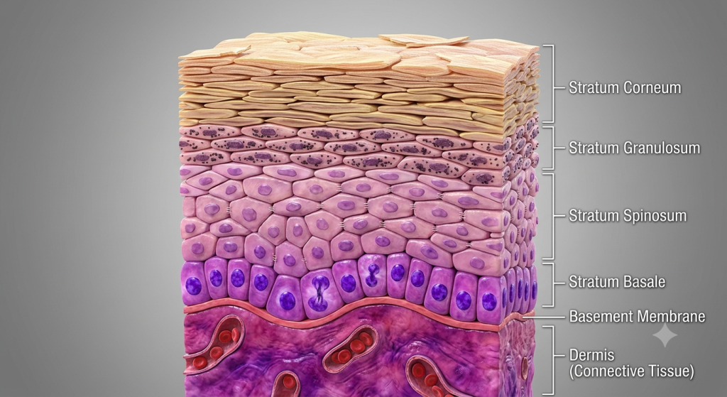
The Stratum Basale (The Base Layer)
This is the deepest layer of the epithelium. It rests directly on a basement membrane, which acts like biological glue holding the tissue to the connective tissue below. Here, the cells look completely different from the surface.
Instead of being flat, these basal cells are plump and cuboidal (cube-shaped) or columnar (column-shaped). They are the hardest workers in the entire tissue. This layer is highly active, constantly undergoing mitosis (cell division) to produce new cells.
The Stratum Spinosum (The Prickly Layer)
Just above the basal layer sits the stratum spinosum. As newly minted cells are pushed up into this layer, they start to change. Under a microscope, these cells appear to have tiny little spines sticking out of them.
These ‘spines’ are actually cellular connections called desmosomes. Desmosomes act like microscopic spot welds, holding the cells tightly together. This extreme cellular bonding is exactly what gives the tissue its incredible resistance to tearing.
The Stratum Granulosum (The Granular Layer)
As cells get pushed further up, they enter the stratum granulosum. At this stage, the cells start flattening out significantly. They also begin to stockpile massive amounts of proteins.
You will see dark clumps inside them called keratohyalin granules. These granules secrete a lipid-rich (fatty) substance that coats the cells. This creates a waterproof barrier, preventing you from losing all your body’s moisture to the dry air around you.
The Stratum Corneum (The Horny Layer)
Finally, we reach the apical surface, or the very top layer. By the time cells make it here, they are dead. Yes, the entire outer surface of your skin is a graveyard of dead, flattened, nucleus-free cells.
These cells are entirely packed with a tough, fibrous protein called keratin. They overlap like shingles on a roof, creating a tough, protective shield against bacteria, friction, and chemicals. When you scratch your arm, these are the cells that flake off.
3. Keratinized vs. Non-Keratinized Tissue: The Big Divide
Not all stratified squamous epithelium is identical. Your body requires different types of protection depending on the location. Therefore, this tissue comes in two highly specialized flavors: keratinized and non-keratinized.
The primary difference comes down to moisture and the presence of that tough protein, keratin. Let’s look at how they compare.
| Feature | Keratinized Epithelium | Non-Keratinized Epithelium |
|---|---|---|
| Primary Location | Epidermis (outer skin), palms, soles of feet | Inside of mouth, esophagus, vagina, anal canal |
| Top Cell Status | Dead, completely flattened, lacking a nucleus | Alive, flattened, retain their nuclei |
| Surface Environment | Dry, exposed to air and severe friction | Moist, coated in mucus or glandular secretions |
| Keratin Levels | Extremely high (packed with tough proteins) | Very low or entirely absent |
The keratinized epithelium is built for absolute war against the outside world. It handles the dry air, ultraviolet radiation, and rough surfaces we encounter daily. The dead surface cells sacrifice themselves to protect the living tissue underneath.
On the flip side, the non-keratinized tissue handles a different kind of stress. Think about swallowing a sharp potato chip. The lining of your esophagus needs to be thick and multi-layered to prevent that chip from slicing into your throat. However, because it stays moist internally, the top cells don’t need to die and pack themselves with dry keratin.
4. The Incredible Journey: Epithelial Regeneration
One of the most mind-blowing facts about stratified squamous epithelium is its regeneration rate. This tissue is not static. It is a highly dynamic, constantly moving escalator of cellular turnover.
Because the main job of this tissue is protection against abrasion, the top layers are constantly being worn away. Every time you wash your face, chew food, or put on clothes, thousands of cells are sloughed off. To prevent you from wearing away to nothing, the tissue must rebuild itself from the bottom up.
💡 Pro Tip: Exfoliating your skin helps speed up the removal of the dead, top-layer cells in the stratum corneum. However, over-exfoliating can strip away cells faster than the basal layer can regenerate them, leading to redness and severe irritation.
The process starts in the stratum basale. Here, stem cells undergo continuous division. When a stem cell divides into two, one daughter cell stays behind to keep dividing later. The other daughter cell gets pushed upward.
As more and more cells are born at the base, the older cells are forced higher into the stratum spinosum, then the granulosum. As they get further away from the nutrient-rich blood supply at the base, they slowly starve and die, eventually flattening out at the top.
According to recent histological cell-tracking data, the complete journey for a skin cell—from its birth in the basal layer to its death and shedding at the surface—takes roughly 28 to 40 days in a healthy human adult.
5. Why This Tissue is the Ultimate Protection Barrier
We’ve talked about the structure, but let’s dive into exactly how this tissue functions as a tissue protection barrier. It defends your body on multiple fronts simultaneously.
Mechanical Defense
The sheer number of cell layers is the first line of defense. A simple epithelium (one layer thick) would tear immediately under stress. The stratified nature means that even if a dozen layers are scraped off by a rough surface, the underlying connective tissue, nerves, and blood vessels remain completely untouched.
Waterproofing and Hydration Control
You are mostly made of water. If your internal environment were directly exposed to the dry air, you would dehydrate rapidly. The lipid-rich secretions in the granular layer, combined with the dense keratin at the top, create a nearly impermeable waterproof seal.
Pathogen Blocking
Bacteria and viruses are constantly trying to invade your body to find a warm, nutrient-rich place to multiply. The tight junctions and overlapping dead cells of the stratum corneum make it incredibly difficult for microbes to squeeze through. Furthermore, as the top layers constantly shed, any attached bacteria are literally thrown away.
6. Animal Epidermis: How Evolution Perfected the Shield
Humans aren’t the only ones relying on this incredible barrier. When we look at animal epidermis across the animal kingdom, we see how evolution has tweaked stratified squamous epithelium to survive extreme environments.
Consider the mighty rhinoceros. Its skin is exceptionally thick, heavily highly keratinized stratified squamous epithelium. The keratin layers are massively multiplied to protect the animal from thorny bushes, sharp branches, and attacks from predators in the savanna.
On the opposite end of the spectrum, look at amphibians like frogs. Frogs actually breathe partially through their skin. Because gases need to pass through the tissue, a thick layer of dead keratinized cells would suffocate them. Therefore, amphibian skin is heavily non-keratinized and very thin, relying on mucus glands to maintain moisture and prevent tearing.
Whales and dolphins present another unique case. Living constantly in water, their epidermal turnover rate is incredibly fast. They shed vast amounts of superficial squamous cells daily to stay streamlined and prevent marine parasites from taking hold on their skin.
7. What Happens When the Barrier Fails?
Like any biological system, the stratified squamous epithelium can experience errors. When the delicate balance of cellular regeneration and shedding is disrupted, various diseases and conditions can emerge. Here’s a look at common issues that target this specific tissue.
| Condition | What Goes Wrong in the Epithelium | Visible Result |
|---|---|---|
| Psoriasis | Basal cells divide far too rapidly. Cells reach the surface in 3-5 days instead of 28 days. | Thick, red, scaly plaques that flake off excessively. |
| Squamous Cell Carcinoma | DNA damage (usually from UV light) causes uncontrolled multiplication of squamous cells. | A persistent, non-healing ulcer or abnormal bump on the skin. |
| Human Papillomavirus (HPV) | The virus infects the basal stem cells, altering their growth signals. | Warts form as the infected cells multiply and thicken the epidermal layers. |
| Calluses | Localized, repeated mechanical friction triggers extra basal cell division as a defense mechanism. | A thickened, hardened patch of heavy keratin on hands or feet. |
💡 Pro Tip: If you notice an area of your skin acting strangely—like an ulcer that won’t heal or a mole that changes shape—you should always consult a dermatologist. Squamous cell carcinoma is highly treatable if caught early, before the abnormal cells break through the basement membrane.
8. Step-by-Step: How the Epithelium Heals a Cut
We know how the tissue regenerates normally. But what happens when you get a deep paper cut that slices right through the stratified cell layers? The repair process is a masterclass in biological engineering. Here is how your body handles the emergency.
- The Immediate Clot: Blood rushes in from the damaged vessels beneath the basement membrane. Platelets quickly clump together, forming a temporary plug to stop the bleeding and block immediate bacterial entry.
- Inflammation & Clean Up: The area swells as white blood cells flood the zone. They act as biological garbage trucks, eating up invading bacteria and dead cellular debris.
- Basal Cell Migration: The surviving stem cells in the stratum basale at the edge of the cut suddenly stop pushing upward. Instead, they flatten out and physically migrate sideways across the wound gap to form a new foundation.
- Rapid Proliferation: Once the basal cells connect across the gap, they kick into overdrive. They divide rapidly, stacking new layers upward to rebuild the spinous, granular, and corneum layers.
- Scab Sloughing: As the new stratified squamous epithelium finishes rebuilding beneath, the dried blood clot (scab) on the surface loses its attachment and naturally falls off, revealing fresh pink tissue underneath.
Clinical wound healing studies show that in an optimal, moist environment, basal epithelial cells can migrate across a wound bed at a rate of roughly 0.5 millimeters per day.
9. Pro Tips for Protecting Your Epithelial Layers
Your stratified squamous epithelium works tirelessly to protect you. It only makes sense that you should put a little effort into protecting it back. Here are some actionable steps you can take to maintain the integrity of your biological armor.
💡 Pro Tip: Maintain Hydration from the Inside Out. Your skin cells need water to function correctly. While lotions help seal moisture in, you must drink enough water daily to supply the basal layers via your bloodstream. Dehydrated tissue becomes brittle and prone to micro-tears.
💡 Pro Tip: Don’t Strip the Lipids. The stratum granulosum provides essential lipids that waterproof your skin. Using extremely harsh soaps or taking scalding hot showers literally melts and strips these natural oils away, compromising your barrier function.
💡 Pro Tip: UV Protection is Non-Negotiable. Ultraviolet radiation from the sun penetrates the top dead layers and damages the DNA of the living basal cells. Always apply broad-spectrum sunscreen to protect the structural integrity of your cellular regeneration process.
10. Frequently Asked Questions (FAQ)
We often get lots of specific questions regarding skin histology and tissue function. Here are some of the most common queries people search for.
Where is stratified squamous epithelium found?
You will find the keratinized version forming the epidermis (your outer skin). The non-keratinized version lines moist internal surfaces subjected to wear and tear, like your oral cavity (mouth), esophagus, vocal cords, vagina, and anal canal.
Why are the top cells flat?
As cells get pushed away from the blood supply at the base, they lose water and nutrients. They slowly die and flatten out into scale-like shapes. This flat shape allows them to overlap tightly, creating a superior, seamless physical barrier.
Can non-keratinized tissue become keratinized?
Yes, under severe stress! If a non-keratinized area (like the inside of your cheek) is constantly subjected to intense friction or chronic irritation, it can undergo a process called metaplasia and start producing keratin to protect itself.
Does this tissue have blood vessels?
No, epithelial tissue is entirely avascular. It does not contain its own blood vessels. The living cells in the lower layers rely on oxygen and nutrients diffusing up from the blood vessels located in the underlying connective tissue.
How does aging affect this tissue?
As we age, the basal stem cells slow down their division rate. This means the epidermis becomes physically thinner. Additionally, it takes much longer to heal from cuts, and the tissue becomes more susceptible to damage from everyday friction.
11. Wrapping Up Our Microscopic Journey
We have covered a massive amount of ground today, looking deep into the hidden microscopic structures that keep us safe. From the rapidly dividing basal cells to the tough, dead, keratin-packed scales on the surface, your stratified squamous epithelium is nothing short of an engineering marvel. It is the ultimate protective barrier, perfectly adapted to shield your delicate internal systems from mechanical stress, dehydration, and hostile invaders.
Understanding your skin histology doesn’t just help you ace a biology test; it gives you the knowledge to take better care of your body’s most critical defense system. By staying hydrated, protecting against UV damage, and respecting your skin’s natural regeneration cycle, you ensure your biological armor stays strong for decades to come.
We love hearing your thoughts on human biology! What surprised you the most about how your skin heals itself after a cut? Drop your thoughts, questions, or your own skincare routines in the comments section below!