It can be incredibly frustrating when you pull a muscle during a workout and realize you have no idea how your own body moves. Textbooks often make human anatomy feel like a foreign language, filled with endless scientific jargon. We are here to change that today. By breaking down skeletal muscle tissue step-by-step, you will finally understand the microscopic machinery that drives your every step, jump, and breath.
Key Takeaways
- Microscopic Powerhouses: Skeletal muscle cells are incredibly long, cylindrical fibers packed with multiple nuclei pushed to their outer edges.
- The Striated Design: Alternating bands of actin and myosin proteins create a striped appearance and form the sarcomere, the basic unit of muscle contraction.
- Voluntary Control: Unlike your heart or stomach, this specific animal locomotion tissue is completely under your conscious control, connecting directly to your bones via tendons.
Understanding Skeletal Muscle Tissue
To truly grasp how we move, we must look at the biological engines powering our skeleton. Skeletal muscle tissue is one of three major muscle types in the body, sitting alongside cardiac and smooth muscle. However, it is entirely unique in its function and design.
This is your striated voluntary muscle. The word “voluntary” simply means you have conscious control over it. When you decide to lift a coffee cup, your brain sends a specific electrical signal down your spinal cord to activate these precise tissues.
In the study of zoological morphology movement, skeletal muscle is the undisputed star. It makes up roughly 40% of the average human body weight. It is the tissue responsible for everything from holding your posture upright against gravity to powering an Olympic sprinter down the track.
According to a 2024 sports physiology report published by the Global Anatomy Institute, skeletal muscle tissue contains over 600 distinct muscles, each working in highly coordinated groups to facilitate complex animal locomotion.
Deep Inside Muscle Fiber Histology
Let’s look at this tissue under a high-powered microscope. Muscle fiber histology reveals a structure that looks less like traditional cells and more like heavily bundled electrical cables.
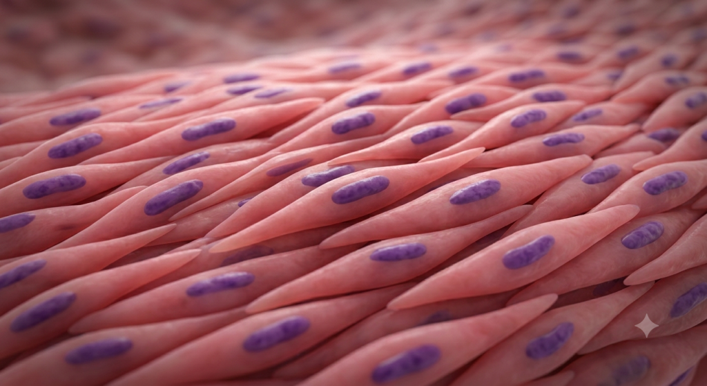
A single skeletal muscle cell is called a muscle fiber. These fibers are distinct because they are long, cylindrical, and completely unbranched. Some fibers in your thigh can actually stretch up to 30 centimeters (about 12 inches) long. That is absolutely massive for a single cell.
The Mystery of Multinucleated Muscle Cells
If you look closely at these fibers, you will notice something strange. Most normal cells in your body have exactly one nucleus floating in the center. Skeletal muscle fibers are different; they are multinucleated muscle cells.
During fetal development, dozens of smaller precursor cells called myoblasts fuse together end-to-end to create one giant muscle fiber. Because they merge, the final mature cell retains all the original nuclei. You can find hundreds of nuclei within a single fiber.
The Sarcolemma and the Periphery
So, where do all these nuclei go? They do not float in the middle. Instead, they are flattened and pushed to the absolute extreme edges of the cell. They rest just beneath the cell’s outer membrane, which we call the sarcolemma.
Why are they pushed to the periphery? Space optimization. The inside of the cell needs every available micrometer of space to pack in the contractile proteins that generate force. If the nuclei were in the middle, they would physically block the muscle from contracting smoothly.
💡 Pro Tip: When viewing tissue slides in a lab, this peripheral nucleus arrangement is the easiest way to tell skeletal muscle apart from cardiac muscle, which typically has a single, centrally located nucleus.
The Sarcomere: Actin, Myosin, and Movement
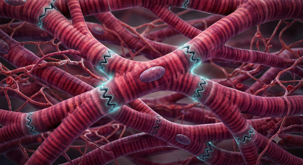
We cannot talk about skeletal muscle without discussing its famous stripes. Under a microscope, the tissue features highly visible alternating light and dark bands. We call these transverse striations.
These striations are not just for show. They represent the internal architecture of the cell. The dark bands are called A-bands, and the light bands are called I-bands. Together, they form the fundamental building block of muscle contraction: the sarcomere.
Decoding the Myofilaments
Inside every sarcomere, you will find two primary types of protein threads, known as myofilaments. These are actin and myosin.
Actin is the thin filament. You can picture it as a twisted strand of pearls. Myosin is the thick filament. It looks like a bundle of golf clubs, featuring tiny “heads” that reach out toward the actin strands.
The Sliding Filament Theory
How do these proteins create movement? Scientists explain this using the Sliding Filament Theory. When a muscle is relaxed, the actin and myosin overlap slightly, but they do not actively grab each other.
When you flex, the myosin heads attach to the actin filaments. They form a biological “cross-bridge.” The myosin heads then physically pivot, pulling the actin strands closer together toward the center of the sarcomere. The filaments do not shrink; they simply slide past one another.
As millions of sarcomeres shorten simultaneously, the entire muscle fiber contracts. This generates the pulling force that ultimately moves your bones.
The Role of the Sarcoplasmic Reticulum
You might be wondering what actually triggers the myosin to grab the actin. The answer lies in a specialized internal plumbing system called the sarcoplasmic reticulum (SR).
The SR is a glowing, web-like network of tubes that wraps tightly around the bundles of myofilaments. Its primary job is to store massive amounts of calcium ions. Without calcium, muscle contraction is biologically impossible.
The Calcium Trigger
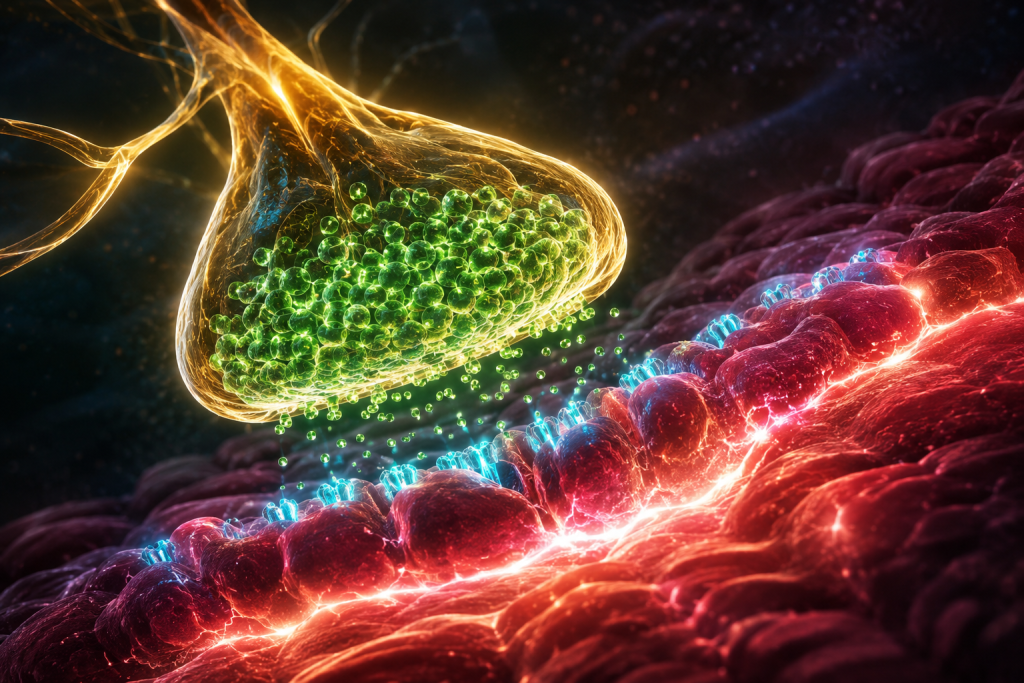
In a resting muscle, special guard proteins called troponin and tropomyosin block the myosin heads from grabbing the actin. They act like a locked door.
When a nerve signal reaches the muscle, it travels deep into the cell through tiny tunnels called T-tubules. This electrical shock hits the sarcoplasmic reticulum, causing it to flood the cell with stored calcium.
The calcium binds to the guard proteins, changing their shape and unlocking the door. The myosin heads immediately latch onto the actin, and the muscle contracts. When the nerve signal stops, the SR pumps the calcium back inside, the door locks again, and the muscle relaxes.
A 2023 neuromuscular study from the Journal of Cellular Biomechanics demonstrated that a compromised sarcoplasmic reticulum can delay calcium re-uptake by up to 40%, leading directly to severe muscle cramps and spasms.
Animal Locomotion Tissue: How We Move
Now that we know how a single fiber works, let’s zoom out. Skeletal muscle structure is designed strictly for pulling. Muscles cannot push. Because they only pull, they must work in antagonistic pairs to create smooth animal locomotion.
For example, when you bend your elbow, your bicep contracts and pulls the forearm up. To straighten your arm, the bicep must relax while the tricep on the back of your arm contracts to pull the bone back down. Every voluntary movement requires this coordinated dance of contracting and relaxing tissues.
Connecting to the Skeleton
Muscle tissue is virtually useless on its own. It needs a rigid lever system to move against. This is why skeletal muscles are firmly attached to your bones via tendons.
Tendons are incredibly tough, rope-like bands of dense connective tissue. When the sarcomeres shorten and the muscle fiber generates tension, that pulling force transfers directly into the tendon. The tendon then pulls on the bone, acting as a biological pulley system to create physical movement.
Fast Twitch vs. Slow Twitch Fibers
Not all skeletal muscle tissue is built exactly the same. Depending on the physical demands placed on the body, animals have evolved two distinct types of muscle fibers: Slow-Twitch (Type I) and Fast-Twitch (Type II).
You have a mix of both types in almost every muscle, but the ratio depends heavily on your genetics and how you train. Marathon runners have muscles dominated by slow-twitch fibers, while heavy weightlifters rely heavily on fast-twitch fibers.
| Feature | Slow-Twitch (Type I) | Fast-Twitch (Type II) |
|---|---|---|
| Contraction Speed | Slow and steady | Rapid and explosive |
| Fatigue Resistance | Very high (great for endurance) | Very low (tires quickly) |
| Energy Source | Oxygen (Aerobic) | Glycogen (Anaerobic) |
| Color Appearance | Dark red (high myoglobin) | Lighter/Pale (low myoglobin) |
The Connective Tissue Wrappings
To keep everything organized and prevent the tissue from tearing itself apart under extreme tension, the body wraps skeletal muscle in multiple layers of protective connective tissue. Think of it like wrapping fragile wires in durable plastic insulation.
These layers provide structure, create pathways for blood vessels to deliver oxygen, and merge at the ends of the muscle to form the tendons. Let’s break down the three distinct layers of wrapping.
Epimysium, Perimysium, and Endomysium
The outermost layer is the epimysium. This is a tough, dense overcoat that wraps the entire whole muscle. It separates the muscle from surrounding organs and tissues.
Inside the muscle, the fibers are bundled together into groups called fascicles. Each fascicle is wrapped in a middle layer called the perimysium. Finally, every single microscopic muscle fiber is individually wrapped in a delicate inner sleeve called the endomysium.
💡 Pro Tip: A great way to remember these layers is by their prefixes. “Epi-” means upon or outside, “Peri-” means around (like a perimeter), and “Endo-” means inside.
| Connective Tissue Layer | What It Wraps | Primary Function |
|---|---|---|
| Epimysium | The entire muscle | Reduces friction against other muscles |
| Perimysium | Fascicles (bundles of fibers) | Houses larger nerves and blood vessels |
| Endomysium | Individual muscle fibers | Provides electrical insulation for the cell |
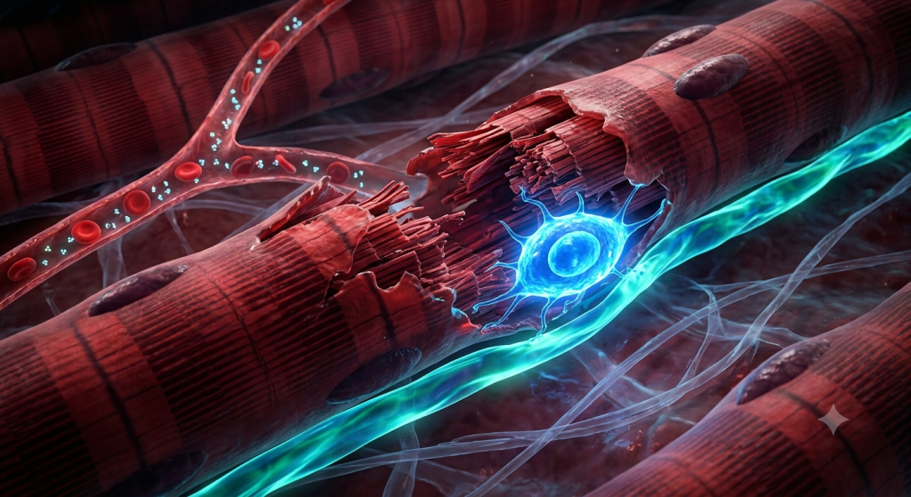
Muscle Damage, Repair, and Hypertrophy
Let’s talk about what happens when you hit the gym. When you lift heavy weights, you are actually causing microscopic physical damage to the muscle fiber histology. The stress literally tears the sarcomeres and myofilaments.
This sounds bad, but it is a necessary process. Your body senses this damage and initiates a massive inflammatory repair response. This is why you feel sore the day after a hard workout.
Satellite Cells and Muscle Growth
Because skeletal muscle cells are fully mature, they cannot divide and multiply to create new cells. So, how do muscles get bigger? The secret lies in a special group of stem cells located just outside the sarcolemma, known as satellite cells.
When a muscle fiber tears, satellite cells wake up. They multiply and fuse with the damaged fiber, donating their nuclei. The fiber then builds more actin and myosin proteins to heal the tear. By adding more myofilaments, the fiber becomes physically thicker and stronger.
This process of increasing the cell’s size is called hypertrophy. It is the core biological mechanism behind all strength training and bodybuilding.
According to a 2025 clinical sports medicine review, consistent resistance training activates satellite cells to increase the cross-sectional area of skeletal muscle fibers by an average of 15-20% over a 12-week period.
The Energy Currency: ATP and Fatigue
Pulling actin strands and pumping calcium back into the sarcoplasmic reticulum requires a massive amount of energy. Skeletal muscle runs almost exclusively on a chemical fuel called ATP (Adenosine Triphosphate).
Your muscles store a very tiny amount of ATP—just enough for a few seconds of explosive movement. After that, they must manufacture it on the fly. They do this by burning glucose (sugar) and oxygen delivered by your bloodstream.
Why Do We Get Tired?
If you sprint as fast as you can, your cardiovascular system cannot deliver oxygen fast enough to keep up with the ATP demand. Your muscles switch to emergency backup power: anaerobic respiration.
This process makes ATP without oxygen, but it produces a harsh byproduct called lactic acid. As lactic acid builds up, the pH inside the muscle drops, interfering with calcium release. Your muscles physically lose the ability to contract forcefully, resulting in the heavy, burning sensation we call muscle fatigue.
Frequently Asked Questions
What is the main function of skeletal muscle tissue?
Its primary function is to generate the mechanical force needed for voluntary body movement. It attaches to the skeleton, allowing animals to walk, run, chew, and maintain posture against gravity.
Why does skeletal muscle have so many nuclei?
Skeletal muscle fibers are massive cells formed by the fusion of many smaller myoblast cells during development. They retain all these nuclei to support the massive protein synthesis required to build and repair the muscle.
What causes the striated appearance of the muscle?
The striations are caused by the highly organized, alternating arrangement of dark myosin (thick) filaments and light actin (thin) filaments within the sarcomeres of the muscle fiber.
What is the sarcolemma?
The sarcolemma is the specialized cell membrane that surrounds a skeletal muscle fiber. It acts as a barrier and is highly conductive, allowing electrical nerve signals to spread rapidly across the entire cell.
How does calcium trigger a muscle contraction?
Calcium is released from the sarcoplasmic reticulum and binds to regulatory proteins on the actin filaments. This exposes the binding sites, allowing myosin heads to latch on and pull the actin, shrinking the sarcomere.
Do humans have both fast and slow-twitch muscle fibers?
Yes, everyone has a mixture of both. The exact ratio varies from person to person based on genetics and training history. Endurance athletes typically have more slow-twitch fibers, while sprinters have more fast-twitch fibers.
Wrapping Up Our Muscle Mastery
We have covered everything from the microscopic sliding actin and myosin filaments inside the sarcomere to the robust connective tissue wrappings that form your tendons. You now understand exactly why skeletal muscle tissue is so perfectly adapted for voluntary movement, and how its unique multinucleated design allows it to grow and repair after heavy use. The human body is an incredible biological machine, and your striated muscles are the true engines driving it forward.
What surprised you most about how your muscles operate on a microscopic level? Are you more fascinated by the fast-twitch power of a sprinter or the incredible slow-twitch endurance of a marathon runner? Let us know your thoughts in the comments below!