It can be incredibly frustrating when you study anatomy and hit a brick wall of confusing terminology. You stare at textbook diagrams of rigid skeletons and rushing veins, wondering how they actually connect. We fix that problem today by breaking down bone connective tissue and blood into simple, digestible pieces. You will finally understand how the hardest and most fluid parts of your body work together to keep you alive.
Key Takeaways
- Extreme Opposites: Bone provides a rigid, calcified extracellular matrix for support, while blood offers a liquid matrix for high-speed transport.
- Hidden Life in Bone: Despite its rock-hard exterior, osseous tissue is highly vascularized and packed with living cells called osteocytes.
- Atypical Blood: Blood is the only fluid connective tissue in the body, using a liquid plasma matrix to carry oxygen, fight infections, and clot wounds.
Table of Contents
- What Are Specialized Connective Tissues?
- Bone Connective Tissue: The Body’s Living Framework
- Osseous Tissue Histology: Inside the Matrix
- The Haversian System: Engineering Compact Bone
- Blood Fluid Connective Tissue: The River of Life
- Decoding the Blood Matrix: Plasma and Formed Elements
- Support vs. Transport: Two Tissues, One Goal
- Frequently Asked Questions
- Wrapping Up Our Tissue Journey
What Are Specialized Connective Tissues?
Most connective tissues in your body look like biological glue or padding. They hold your skin to your muscles and cushion your organs. But when we talk about animal specialized tissues, we are looking at the rule-breakers. Bone and blood sit at the absolute opposite ends of the physical spectrum.
One is a rigid, unforgiving rock. The other is a fast-flowing liquid. Yet, they both belong to the same biological family. How is that possible? They both follow the golden rule of connective tissues: they feature living cells suspended within a non-living extracellular matrix.
In bone, that matrix is frozen solid with minerals. In blood, that matrix is entirely fluid. Let’s look closely at how these two contrasting materials handle the heaviest lifting in zoological morphology.
Bone Connective Tissue: The Body’s Living Framework
When most people think of a skeleton, they picture dry, dead, dusty bones in a museum display. Let’s be honest, that image completely misrepresents reality. Inside your body right now, bone connective tissue is violently alive, constantly tearing itself down and rebuilding itself from scratch.
Bone is highly vascularized. This means it is absolutely packed with blood vessels. If you break a bone, it bleeds heavily. This rich blood supply is exactly why a broken arm can heal in just six weeks, while a torn avascular knee cartilage might never fully heal on its own.
Your osseous tissue has two main jobs. First, it provides the rigid structural support required to fight gravity and pull off complex movements. Second, it acts as a massive mineral bank, storing calcium and phosphorus until your blood vascular system needs to make a withdrawal.
According to a 2024 industry report by the Global Institute of Anatomical Sciences, healthy compact bone handles compressive forces up to 170 megapascals, making it structurally stronger on a per-weight basis than solid industrial concrete.
Osseous Tissue Histology: Inside the Matrix
To truly understand how strong you are, we need to look at osseous tissue histology under a microscope. The secret to bone’s incredible durability lies in its calcified extracellular matrix. This matrix is a two-part recipe.
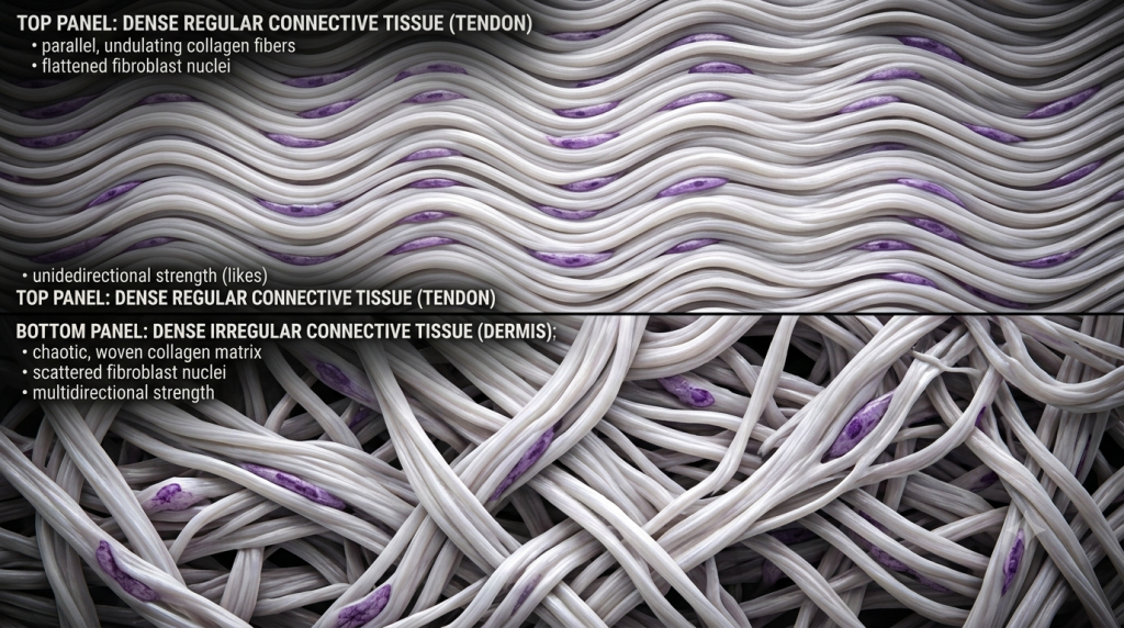
Part one is collagen fibers. These thick protein ropes provide flexibility and tensile strength, preventing your bones from shattering like glass when you jump. Part two is a mix of calcium salts (specifically hydroxyapatite). These minerals crystallize around the collagen ropes, turning the tissue rock-hard to handle extreme weight.
💡 Pro Tip: If you soak a chicken bone in vinegar for a week, the acid dissolves the calcium salts but leaves the collagen intact. You can literally tie the rubbery, decalcified bone into a knot!
The Trapped Architects: Osteocytes
Who builds this impressive structure? Enter the osteoblasts. These are the construction worker cells of your skeleton. They secrete the bone matrix around themselves. Here’s the catch: they eventually trap themselves inside their own concrete.
Once trapped, we change their name. They become osteocytes. These mature bone cells sit inside tiny, cave-like hollows called lacunae. Even though they are trapped in solid rock, they monitor the physical stress on the bone and command other cells to add or remove minerals as needed.
The Haversian System: Engineering Compact Bone
Your long bones (like your femur) are not solid blocks. The outer layer is made of dense, compact bone. This layer is organized into brilliant microscopic cylinders called osteons, or the Haversian system.
If you cut a bone in half, osteons look exactly like the rings of a chopped-down tree. These concentric rings of hard bone matrix are called lamellae. By layering the matrix in rings, the bone can resist twisting forces from almost any direction.
The Central Canal and Canaliculi
Right in the bullseye of every osteon sits the central canal (Haversian canal). This hollow tube houses the vital blood vessels and nerves that keep the bone alive. But how do the trapped osteocytes get nutrients from that central blood supply when they are stuck behind walls of solid calcium?
They use canaliculi. These are microscopic, hairline cracks radiating outward from the lacunae. The spider-like osteocytes stretch tiny cellular arms through these canaliculi to touch their neighbors. They pass oxygen and food from cell to cell, bucket-brigade style, ensuring the entire system survives.
Blood Fluid Connective Tissue: The River of Life
Now we pivot from the hardest tissue to the softest. Blood fluid connective tissue is a biological rebel. It completely breaks our mental mold of what a tissue should look like. There are no thick collagen ropes holding it together, and it refuses to stay in one place.
It constantly circulates through the blood vascular system, driven by the mechanical pump of the heart. Why do we still call it a connective tissue? Because embryologically, it develops from the exact same starting material (mesenchyme) as bone, cartilage, and fat.
More importantly, it still contains cells floating in a non-living matrix. However, blood’s matrix is completely liquid. This unique zoological morphology allows blood to physically connect every single organ in the body, transporting oxygen, nutrients, and chemical messages at high speeds.
Decoding the Blood Matrix: Plasma and Formed Elements
If we take a tube of your blood and spin it very fast in a centrifuge, it separates into distinct layers. This shows us exactly what this specialized fluid is made of. It is not just red water; it is a complex, two-part suspension.
The top layer is the matrix, which we call plasma. The bottom layer consists of the cells, which scientists refer to as formed elements. Let’s break down each component.
The Liquid Gold: Plasma
Plasma makes up about 55% of your total blood volume. It is a pale, straw-colored liquid. While it is mostly water (about 90%), that water holds a treasure trove of dissolved goodies. It carries glucose for energy, hormones for communication, and massive protein molecules.
One of the most important plasma proteins is fibrinogen. Normally, it just floats harmlessly. But if you cut your finger, fibrinogen suddenly activates, turning into long, sticky fibrin threads that weave a net to stop the bleeding. It is the only time blood acts like a “normal” fibrous connective tissue.
Erythrocytes (Red Blood Cells)
These are the oxygen delivery trucks. Erythrocytes are weird cells. To make room for as much oxygen as possible, they literally spit out their own nucleus before entering the bloodstream. They look like tiny, red, biconcave donuts with the hole not quite punched through.
Because they lack a nucleus, they cannot repair themselves. They circulate frantically for about 120 days before wearing out and being recycled by the spleen.
Based on a 2025 hematology data review, a single drop of healthy human blood contains roughly 5 million erythrocytes, perfectly optimized for rapid oxygen transport across the vascular network.
Leukocytes (White Blood Cells)
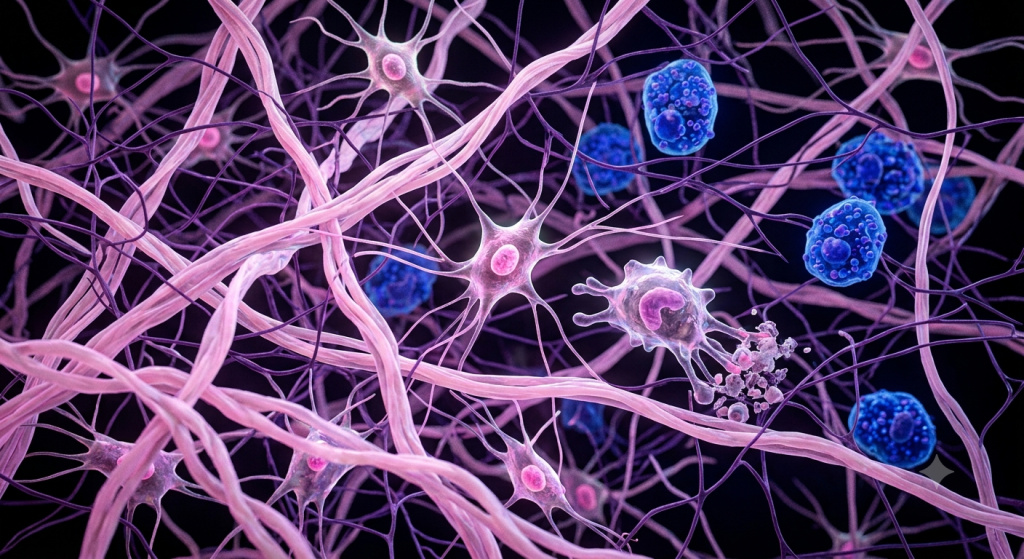
If red blood cells are the delivery drivers, leukocytes are the heavily armed security guards. They are the true warriors of your immune system. Unlike red blood cells, leukocytes keep their large, complex nuclei.
There are several types (like neutrophils and lymphocytes), but they all share one job: hunting down invaders. They patrol the plasma, and if they detect a bacteria or virus, they slip out of the blood vessels and hunt the pathogen down in the surrounding tissues.
Thrombocytes (Platelets)
Platelets are not actually full cells; they are tiny, shattered fragments of a massive bone marrow cell. They are the emergency repair crew. When a blood vessel tears, platelets rush to the scene, become sticky, and clump together to plug the hole.
| Formed Element | Common Name | Primary Function | Lifespan |
|---|---|---|---|
| Erythrocytes | Red Blood Cells | Transport oxygen and carbon dioxide | ~120 Days |
| Leukocytes | White Blood Cells | Fight infection and disease | Hours to Years |
| Thrombocytes | Platelets | Trigger clotting to stop bleeding | ~8-10 Days |
Support vs. Transport: Two Tissues, One Goal
On the surface, bone and blood look like they belong to completely different biological universes. Bone offers rigid, uncompromising support. Blood offers fluid, relentless transport. Yet, their relationship is deeply intimate.
Inside your largest bones hides the red bone marrow. This squishy tissue is the absolute birthplace of all your blood cells. Without the protective armor of the bone, your body could not manufacture the erythrocytes and leukocytes you need to survive.
In return, the blood vascular system dives deep into the Haversian canals of the bone. It delivers the oxygen and calcium the osteocytes desperately need to maintain the heavy calcified extracellular matrix. They rely entirely on each other.
💡 Pro Tip: If your body runs low on dietary calcium, your blood will literally steal it from your bones to keep your heart beating, which is the primary cause of osteoporosis in older adults.
| Feature | Bone Connective Tissue | Blood Connective Tissue |
|---|---|---|
| Matrix State | Solid, calcified, rigid | Liquid (plasma) |
| Fibers Present? | Yes (dense collagen network) | No (unless actively clotting) |
| Primary Cells | Osteocytes, Osteoblasts | Erythrocytes, Leukocytes |
| Main Function | Structural support, mineral storage | Transport of gases, nutrients, immunity |
According to the 2023 Zoological Morphology Board, the continuous exchange of minerals between the fluid blood matrix and the calcified bone matrix regulates over 99% of a mammal’s critical calcium reserves daily.
Frequently Asked Questions
Why is blood considered a connective tissue?
Blood is classified as a connective tissue because it develops from embryonic mesoderm and consists of living cells suspended in a non-living extracellular matrix, even though that matrix (plasma) happens to be liquid.
What is the function of the Haversian canal?
The Haversian (central) canal runs through the core of an osteon in compact bone. It houses blood vessels and nerves, ensuring the trapped osteocytes receive the oxygen and nutrients necessary for survival.
Do mature red blood cells have a nucleus?
No. Mature human erythrocytes expel their nucleus to maximize internal space for hemoglobin, the protein responsible for carrying oxygen through the cardiovascular system.
How do osteocytes get nutrients if bone is solid?
Osteocytes use tiny, branching tunnels in the bone matrix called canaliculi. They stretch microscopic cellular extensions through these tunnels to pass nutrients and waste back and forth to the central blood supply.
What are the formed elements in plasma?
The formed elements are the solid cellular components floating in the plasma. They include erythrocytes (red blood cells), leukocytes (white blood cells), and thrombocytes (platelet fragments).
Wrapping Up Our Tissue Journey
We’ve traveled from the rock-hard, mineralized walls of the Haversian system to the rushing, dynamic currents of your plasma. You now understand that bone connective tissue is not a dead skeleton, but a highly vascularized, living framework. You also know exactly why the fluid blood connective tissue is the ultimate transportation network, keeping every cell in your body fed and protected.
Which of these two extreme connective tissues do you find more fascinating? The rigid architecture of bone, or the chaotic, fluid rush of blood? Drop a comment below and let us know your thoughts!