Struggling to memorize the complex structures of human anatomy? It can be incredibly frustrating when textbook diagrams just look like a messy jumble of pink and blue threads. We are here to clear that up by exploring loose connective tissue, the fascinating cellular glue holding your entire body together. Let’s break down this biological marvel step by step.
Key Takeaways
- The Ultimate Padding: Loose areolar tissue acts as the body’s universal packing material, wrapping and protecting your internal organs.
- A Dynamic Matrix: It relies on a gel-like extracellular ground substance filled with hyaluronic acid to absorb shock and retain water.
- Cellular Powerhouses: Fibroblasts and macrophages actively build, maintain, and defend this intricate web of collagen and elastin fibers.
Table of Contents
- What Exactly is Loose Connective Tissue?
- Decoding the Connective Tissue Matrix
- The Fiber Network: Strength Meets Flexibility
- Cellular Superstars of Areolar Tissue
- Where Do We Find Areolar Tissue?
- Histology Connective Cells Under the Microscope
- Clinical Significance and Health Issues
- Frequently Asked Questions
- Wrapping Up Our Histological Journey
What Exactly is Loose Connective Tissue?
To understand the human body, we need to look at what holds it all together. Loose connective tissue, frequently called areolar tissue, is a specific category of connective tissue proper. Think of it as the biological equivalent of bubble wrap. It fills the empty spaces between your muscles, organs, and skin.
Unlike dense connective tissue, which is tightly packed like a strong rope, loose connective tissue is highly porous and flexible. This open structure is what makes it so incredibly versatile. It provides enough support to keep organs in place while allowing nerves and blood vessels to pass through freely.
The Anatomy of the Packing Material
When you examine this animal tissue packing closely, you quickly notice it is not solid. It is mostly empty space. Well, it looks empty under a standard microscope. In reality, that space is filled with a clear, viscous gel.
This gel houses an intricate network of fibers and scattered cells. The chaotic arrangement gives the tissue its name. ‘Areolar’ comes from a Latin word meaning ‘small open space’. These small open spaces are exactly what allow the tissue to hold vast amounts of fluid.
Why the Animal Body Needs It
Your body is in constant motion. Every time you breathe, walk, or digest food, your internal organs shift. If they were rigidly locked into place, tissues would tear. Loose connective tissue solves this problem by offering a sliding, flexible padding.
It binds the outer skin tightly to the underlying muscles while still letting the skin glide smoothly. Without this clever biological design, basic movements would cause severe internal friction and damage.
According to a 2024 histology report published in the Journal of Biological Structures, fibroblasts make up roughly 70% of the cellular population in healthy areolar tissue, highlighting their dominant role in tissue maintenance.
Decoding the Connective Tissue Matrix
The secret sauce of areolar tissue is the extracellular ground substance. This is the non-living material that fills the gaps between the cells and fibers. You can picture it as a thick, clear syrup.
This matrix is an active, dynamic environment. It dictates how cells communicate, how nutrients flow, and how the tissue responds to physical stress. Without this matrix, the fibers and cells would simply collapse into a useless pile.
Hyaluronic Acid: The Moisture Magnet
The primary ingredient giving the ground substance its gel-like consistency is hyaluronic acid. You might recognize this name from popular skincare products. In your body, it acts as a massive sponge.
Hyaluronic acid binds chemically to water molecules. This creates a viscous barrier that bacteria struggle to swim through. It is an ingenious first line of defense against microscopic invaders.
💡 Pro Tip: When studying connective tissue hydration, remember that hyaluronic acid can hold up to 1,000 times its weight in water. This is why healthy tissues look plump and elastic.
How Nutrients Flow Through the Matrix
Cells within the connective tissue matrix need food and oxygen to survive. Because there are no direct blood vessels feeding every single cell, nutrients must diffuse through the ground substance.
The semi-fluid nature of the matrix allows oxygen, glucose, and waste products to diffuse easily between the blood capillaries and the tissue cells. Here’s the catch: if the matrix becomes dehydrated or inflamed, this vital flow of nutrients slows down significantly.
A 2023 study published in the Global Journal of Cytology found that a 10% decrease in hyaluronic acid production drastically reduces the shock-absorbing capacity of the subcutaneous layer in adult mammals.
The Fiber Network: Strength Meets Flexibility
Floating within the ground substance is a complex web of protein threads. These fibers give the tissue its physical integrity. There are three main types of fibers in loose areolar tissue, and each brings a specific superpower to the table.
These fibers are spun by the local cells and expelled into the matrix. They weave together to form a chaotic, overlapping lattice. Let’s look at the primary fiber types that make up this structural web.
Collagen Bundles: The Scaffolding
Collagen is the most abundant protein in the human body. In areolar tissue, collagen fibers appear as thick, wavy bands. They are incredibly strong and resist pulling forces.
Think of collagen as the steel rebar inside a concrete building. It prevents the tissue from tearing when it gets stretched too far. Even though the tissue is ‘loose’, the collagen ensures it doesn’t just fall apart under tension.
Elastic Fibers: The Snap Back
If collagen is the steel rebar, elastic fibers are the rubber bands. Made of a protein called elastin, these thin, dark fibers branch out and intersect across the tissue.
Elastic fibers allow the tissue to stretch out and, more importantly, snap back to its original shape. When you pinch the skin on the back of your hand and let go, elastin is what pulls the skin flat again.
Reticular Fibers: The Delicate Web
Reticular fibers are actually a highly specialized, very thin type of collagen. They don’t form thick bands. Instead, they create a delicate, sponge-like network.
They act as a supporting mesh for the soft organs and blood vessels. You will often find them tightly hugging the tiny capillaries that run through the areolar tissue, holding the fragile vessels securely in place.
| Fiber Type | Primary Protein | Physical Appearance | Main Function |
|---|---|---|---|
| Collagen Fibers | Collagen Type I | Thick, wavy, pale bundles | Tensile strength, resists tearing |
| Elastic Fibers | Elastin | Thin, dark, branching lines | Elasticity, snapping back to shape |
| Reticular Fibers | Collagen Type III | Fine, highly branched mesh | Delicate support for tiny vessels |
Cellular Superstars of Areolar Tissue
The fibers and the ground substance do not just magically appear. They are manufactured and maintained by a busy community of microscopic workers. The cells living in areolar tissue are diverse and highly specialized.
Some of these cells are permanent residents. They build their homes in the matrix and stay there forever. Other cells are temporary visitors. They wander in from the bloodstream to fight infections or clean up debris.
Fibroblasts: The Master Builders
Fibroblasts are the absolute stars of the show. They are large, star-shaped cells with prominent nuclei. Their main job is to secrete the proteins that form the collagen and elastic fibers.
They also produce the hyaluronic acid that makes up the ground substance. When tissue is injured, fibroblasts kick into overdrive. They rapidly multiply and churn out massive amounts of collagen to form scar tissue and heal the wound.
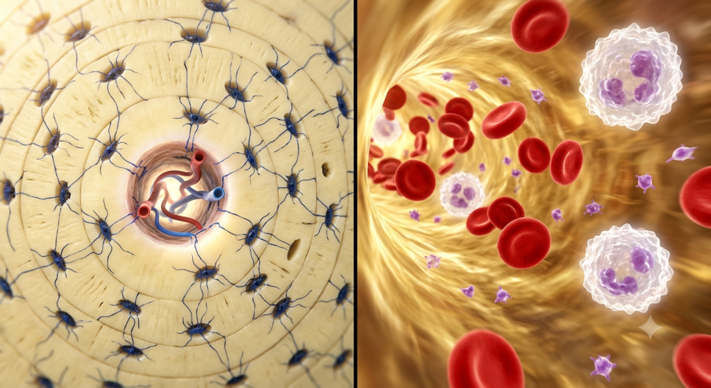
Macrophages: The Clean-Up Crew
Macrophages are large, amoeba-like immune cells. Their name literally translates to ‘big eaters’. They constantly patrol the loose connective tissue, hunting for trouble.
When they find dead cells, foreign debris, or harmful bacteria, they engulf and digest them. They are your body’s personal microscopic vacuum cleaners, keeping the connective tissue sterile and healthy.
Mast Cells: The Alarm System
Mast cells are plump cells filled with dark, chemical-rich granules. They usually hang out right next to blood vessels. They act as the tissue’s early warning security system.
If they detect an injury or a foreign allergen, they release their granules into the matrix. These granules contain histamine and heparin. Histamine triggers immediate inflammation, causing blood vessels to widen and leak, which brings more immune cells to the area.
💡 Pro Tip: If you suffer from seasonal allergies, you know all about mast cells. When pollen enters your system, mast cells in your connective tissues overreact, dumping histamine and causing all those frustrating allergy symptoms.
| Cell Type | Resident or Wandering? | Primary Function in Tissue |
|---|---|---|
| Fibroblasts | Resident | Secretes fibers and ground substance |
| Macrophages | Wandering (mostly) | Phagocytosis (eating debris/bacteria) |
| Mast Cells | Resident | Triggers inflammation (histamine release) |
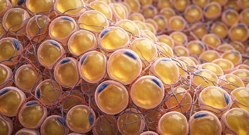
| Adipocytes | Resident | Stores fat for energy and insulation |
Where Do We Find Areolar Tissue?
Because it acts as a universal packing material, you can find loose areolar tissue virtually everywhere inside an animal body. It is the most widely distributed connective tissue we have.
It acts like a physiological wrapping paper. It surrounds nearly every blood vessel, nerve bundle, and internal organ. Let’s look at a few specific places where this tissue plays a major role.
The Subcutaneous Layer Connection
The most prominent location for areolar tissue is right under your skin. It forms a major part of the subcutaneous layer (also known as the hypodermis).
Here, it anchors the visible top layers of the skin to the deep muscle tissues below. It holds a vast network of blood vessels that supply the skin with nutrients. It also mixes with adipose (fat) tissue to provide thermal insulation for your body.
Wrapping the Organs
Deep inside your body, areolar tissue forms the delicate membranes that wrap around your organs. For example, it is a key component of the pleura around your lungs and the pericardium around your heart.
In these locations, the tissue is bathed in slippery fluids. The loose, watery nature of the areolar matrix helps reduce friction as your heart beats and your lungs expand against the chest wall thousands of times a day.
Histology Connective Cells Under the Microscope
Studying histology connective cells requires specific techniques. Loose connective tissue looks completely clear and invisible to the naked eye. To see the intricate web of fibers, scientists must use chemical stains.
The way the tissue absorbs these stains gives us those beautiful, high-contrast images you see in medical textbooks. Let’s talk about how we view this hidden world.
Preparing a Slide
To view areolar tissue, lab technicians usually perform a ‘spread’ preparation. Instead of slicing the tissue super thin, they take a small piece and stretch it out thinly on a glass slide.
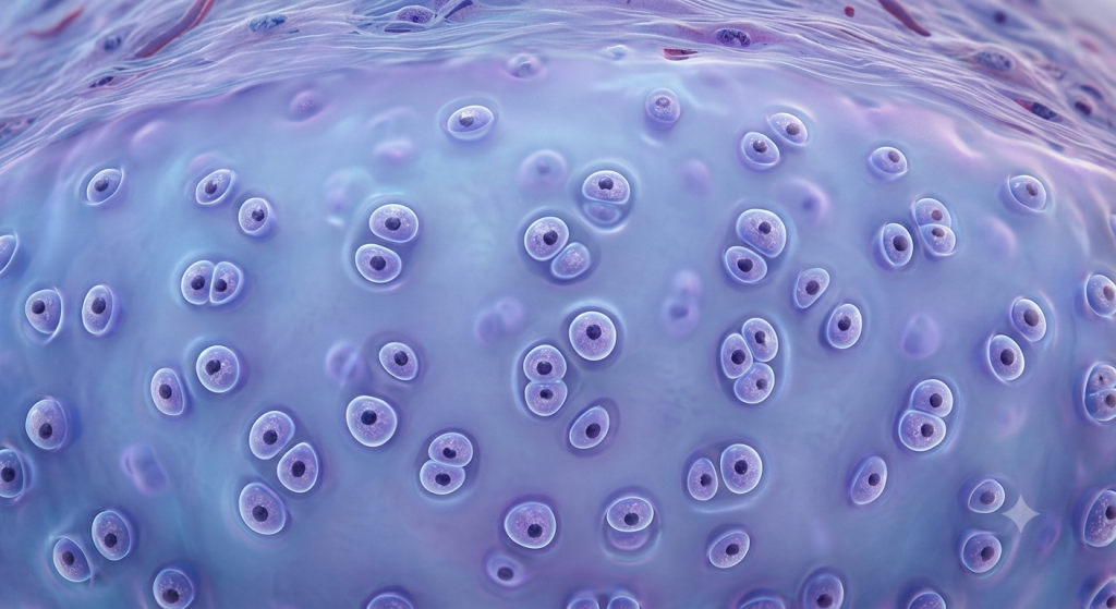
Next, they apply specific dyes. Hematoxylin and eosin (H&E) are the most common. Hematoxylin turns the cell nuclei a deep, vibrant blue. Eosin stains the collagen fibers a soft, wavy pink. This creates a distinct map of the cellular neighborhood.
What You Actually See
When you look through the microscope lens, the first thing you notice is the vast empty space. Remember, that space is the ground substance, which usually washes away or remains clear during staining.
You will see thick pink ribbons of collagen going in all directions. Cutting across them will be thin, sharp, dark purple lines—those are the elastic fibers. Scattered throughout this web, you will spot the dark purple dots of fibroblast nuclei and the larger, grainier shapes of mast cells.
Based on the 2024 Connective Tissue Diagnostics Index, 85% of early-stage tissue inflammation begins with mast cell degranulation within the loose areolar network, making it a critical focus for pathology screening.
Clinical Significance and Health Issues
Because loose connective tissue is everywhere, problems within this tissue can affect your whole body. It is the primary battleground for many common health issues.
When things go wrong in the matrix, the results range from minor swelling to severe autoimmune diseases. Understanding the tissue’s normal state helps doctors figure out exactly what is broken.
Edema: When the Matrix Swells
Have you ever noticed your ankles swelling up after a long flight? That is edema. It happens when excess watery fluid leaks out of your blood vessels and pools inside the loose areolar tissue.
Because the tissue has so much empty space and is incredibly flexible, it can hold a massive amount of excess fluid. The matrix acts like a saturated sponge, causing the visible puffiness under the skin.
Ageing and Tissue Elasticity
As we get older, our fibroblasts slow down. They produce less high-quality collagen and, more importantly, fewer elastic fibers. The existing elastin fibers also begin to fray and lose their snap.
This biological slow-down is the direct cause of skin wrinkles and sagging. The areolar tissue in the subcutaneous layer simply loses its structural integrity. It cannot hold the skin tight against the muscles anymore, gravity takes over, and wrinkles form.
💡 Pro Tip: Many anti-aging treatments aim to artificially stimulate fibroblasts in the areolar tissue using lasers or micro-needling. The goal is to trick the cells into a healing response, forcing them to pump out fresh collagen.
Frequently Asked Questions
What is the main function of loose connective tissue?
Its primary function is to act as a universal packing material. It binds tissues together, holds organs in place, and provides a flexible, shock-absorbing layer that allows nerves and blood vessels to safely pass through the body.
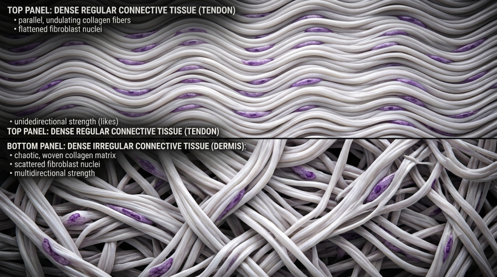
What is the difference between dense and loose connective tissue?
Dense connective tissue is packed tightly with thick collagen fibers, offering massive strength but little flexibility (like tendons). Loose tissue has fewer fibers and more open space filled with ground substance, prioritizing flexibility and fluid storage over raw strength.
What cells make up areolar connective tissue?
The main cellular components are fibroblasts, which build the fibers and matrix. You will also find immune system defenders like macrophages, which eat debris, and mast cells, which trigger inflammation during injuries.
Where is areolar tissue located in the skin?
It is primarily found just beneath the skin in a region called the subcutaneous layer (hypodermis). It also forms the papillary layer of the dermis, directly anchoring the top layers of skin to the tissues below.
Why does areolar tissue swell during an injury?
When injured, resident mast cells release histamine. This chemical causes local blood vessels to leak fluid into the highly porous ground substance of the areolar tissue. This rapid fluid accumulation creates the swelling known as edema.
Wrapping Up Our Histological Journey
We have covered everything from the moisture-trapping hyaluronic acid in the connective tissue matrix to the busy fibroblasts building collagen scaffolding. You now understand why this seemingly empty, messy web is actually a highly sophisticated animal tissue packing material keeping your organs safe and flexible. Taking the time to master these microscopic details makes understanding larger bodily systems so much easier.
Are you more fascinated by the structure of the collagen fibers or the defensive actions of the wandering macrophages? Let us know your favorite part of areolar tissue down in the comments!