You are staring at a microscope slide, and those tissue samples look like a confusing jumble of pink and purple dots. It is incredibly frustrating when you cannot tell one cell layer from another, risking a bad grade or a fundamental misunderstanding of human physiology. We’ve got you covered with this definitive guide to pseudostratified epithelium and transitional epithelium.
Key Takeaways
- Appearance vs. Reality: Pseudostratified tissue looks multi-layered but is actually a single layer attached to one basement membrane.
- Ultimate Stretch: Transitional epithelium (urothelium) is uniquely built to expand and contract, primarily lining the urinary system.
- Location Matters: You will find pseudostratified cells mostly in the respiratory tract, while transitional cells dominate the bladder and ureters.
Table of Contents
- What is Pseudostratified Epithelium?
- The Respiratory Connection
- Introducing Transitional Epithelium
- The Mechanics of Stretching
- Head-to-Head Comparison
- Clinical Significance
- Frequently Asked Questions
- Wrapping Up Our Histology Journey
What is Pseudostratified Epithelium?
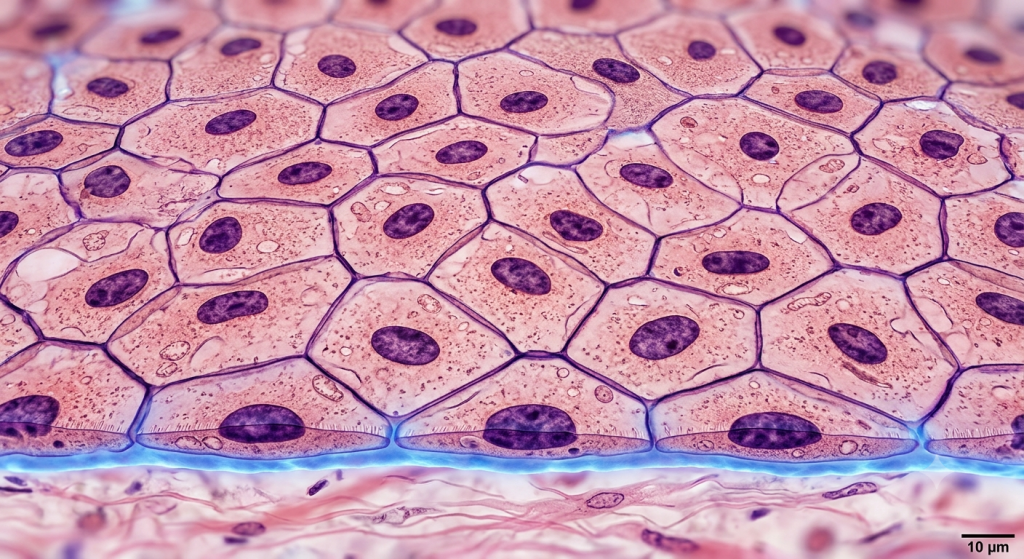
Let’s start by breaking down the word itself. ‘Pseudo’ means false, and ‘stratified’ means layered. So, we are looking at a tissue that fakes being multi-layered. This biological trickery confuses biology students worldwide.
If you examine this tissue under a microscope, you’ll see cellular nuclei scattered at various heights. Some nuclei sit near the bottom, while others rest near the top. This scattered arrangement creates the optical illusion of several distinct cell layers piled on top of one another.
Here’s the catch: every single cell in this tissue actually touches the basement membrane. The basement membrane is the foundational glue holding the tissue to the underlying connective tissue. Because all cells touch the base, it is technically a simple (single-layered) epithelium. Not all cells reach the free apical surface, though. The shorter basal cells act as stem cells, ready to replace taller columnar cells when they die.
According to a 2023 histological survey published in the Global Anatomy Journal, over 85% of early medical students misidentify pseudostratified layers as stratified during their first lab exam.
The Respiratory Connection
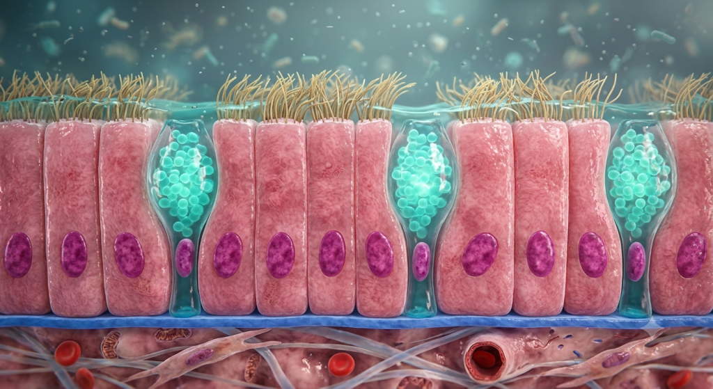
Now, let’s talk about where this sneaky tissue lives. The most famous example is pseudostratified ciliated columnar epithelium. That is a mouthful, but it perfectly describes the lining of your upper respiratory tract.
This specific animal histology type lines your trachea (windpipe) and most of your nasal cavity. The cells here have tiny, hair-like projections called cilia on their top surface. Alongside these ciliated cells, you will find goblet cells. Goblet cells are specialized microscopic factories that pump out mucus.
Together, they form the mucociliary escalator. The goblet cells secrete sticky mucus to trap dust, bacteria, and debris you breathe in. Then, the lush, moving cilia beat upward in a synchronized rhythm. They push the dirty mucus away from your lungs and up toward your throat, where you swallow or cough it out.
💡 Pro Tip: When studying slides of the trachea, always look for the fuzzy border at the top edge of the cells. That fuzziness is the cilia, a dead giveaway that you are looking at respiratory epithelium.
Introducing Transitional Epithelium (Urothelium)
Moving on, we have transitional epithelium. We also call it urothelium tissue because it almost exclusively lines the organs of the urinary system. You will find it in the renal pelvis, ureters, urinary bladder, and parts of the urethra.
Unlike our previous tissue, transitional epithelium is genuinely stratified. It consists of multiple true layers of cells. The most fascinating part about this tissue is its dynamic zoological morphology. It literally changes shape depending on the physical stress applied to it.
The basal cells at the bottom are usually cuboidal or columnar. The intermediate layers consist of polyhedral cells. The surface layer, however, features large, rounded cells known as umbrella cells. These umbrella cells often contain two nuclei and look plump and dome-shaped when the tissue is relaxed.
A 2024 tissue biomechanics report from the Urothelial Research Institute states that healthy transitional epithelium can stretch up to 300% of its resting surface area without tearing.
The Mechanics of Stretching
Think of your urinary bladder like a water balloon. When it is empty, the balloon is thick and small. When you fill it with water, the rubber stretches out, becoming very thin and expanding in volume.
Transitional epithelium does exactly this. When your bladder is empty, the tissue is in its relaxed state. It looks thick, perhaps five to six cell layers deep, with those big dome-shaped umbrella cells bulging at the top.
As the bladder fills with urine, the tissue undergoes severe stretching. The cells slide past one another to accommodate the increased volume. The entire epithelial layer thins out, looking like it’s only two or three layers thick. Those plump umbrella cells flatten out completely, resembling squamous (flat) cells. This incredible elasticity prevents your bladder from bursting and stops toxic urine from leaking into your abdominal cavity.
Head-to-Head Comparison
To fully grasp these complex epithelial layers, we need to compare them side-by-side. Visualizing the differences makes studying much easier.
| Feature | Pseudostratified Epithelium | Transitional Epithelium |
|---|---|---|
| True Layers | Single layer (Simple) | Multiple layers (Stratified) |
| Nuclei Arrangement | Staggered, varying heights | Stacked in distinct cell layers |
| Apical Surface Cells | Often ciliated, columnar | Dome-shaped ‘umbrella’ cells |
| Primary Function | Secretion, mucus transport | Extreme stretching, distension |
On top of their structural differences, their locations and associated structures vary wildly. Let’s look at where they live and what helper cells they rely on.
| Characteristic | Pseudostratified Location | Transitional Location |
|---|---|---|
| Primary Organ System | Respiratory System | Urinary System |
| Specific Organs | Trachea, bronchi, nasal cavity | Bladder, ureters, renal pelvis |
| Specialized Cells Present | Goblet cells (mucus) | Binucleated umbrella cells |
Clinical Significance
Understanding these tissues isn’t just for passing exams. They play massive roles in your daily health. When these tissues fail or mutate, disease quickly follows.
Let’s look at the respiratory tract first. Chronic cigarette smoking wreaks havoc on pseudostratified ciliated tissue. The toxic smoke burns the delicate cilia and forces the tissue to adapt. It changes from a delicate, single-layered ciliated tissue into a tough, multi-layered squamous tissue. We call this process metaplasia.
Without cilia, you can no longer sweep mucus up and out of your lungs. This leads to the infamous ‘smoker’s cough’ as the body violently tries to expel trapped dirt and bacteria.
Based on data from a 2022 respiratory health review, a single smoking-induced metaplasia event can reduce ciliary clearance efficiency in the pseudostratified epithelium by up to 60% within weeks.
For transitional epithelium, urinary tract infections (UTIs) are the most common issue. Bacteria like E. coli have special appendages that allow them to stick specifically to umbrella cells. Once attached, they can invade the tissue and cause painful inflammation. Medical professionals must understand urothelium tissue mechanics to develop better treatments for chronic bladder issues.
💡 Pro Tip: If you are looking at a pathology report mentioning ‘squamous metaplasia’ in a respiratory biopsy, you know the original pseudostratified tissue has been heavily damaged by environmental stressors.
Frequently Asked Questions
Is pseudostratified epithelium simple or stratified?
It is technically a simple epithelium. Even though it looks like multiple layers due to scattered nuclei, every single cell is directly anchored to the bottom basement membrane.
Why is it called transitional epithelium?
Scientists named it ‘transitional’ because they initially thought it was a tissue caught in a transition phase between stratified squamous and stratified columnar tissues. Today, we know it is a distinct, highly elastic tissue type.
Do all pseudostratified cells have cilia?
No, not all of them. While the respiratory tract features ciliated versions, you can find non-ciliated pseudostratified epithelium in parts of the male reproductive system, like the epididymis.
What happens to the bladder umbrella cells when stretched?
When the bladder fills, the dome-shaped umbrella cells flatten out drastically. They change from a plump, cuboidal shape into a thin, squamous-like shape to allow the organ to expand without tearing.
Can damaged transitional epithelium heal?
Yes, urothelium tissue has a very high regenerative capacity. The basal cells at the bottom multiply rapidly to replace damaged or infected surface cells, maintaining the waterproof barrier of the urinary tract.
Wrapping Up Our Histology Journey
We’ve walked through the complex cellular nuclei arrangement of pseudostratified tissue and the incredible tissue elasticity of transitional epithelium. You now understand how to spot the false layers in your airway tissues and how the umbrella cells protect your bladder from bursting under pressure. Mastering these concepts will make your next lab session a breeze.
Which of these two tissue types do you find more fascinating structurally? Drop a comment below and let us know your thoughts!