Are you staring at histology slides, totally lost on how to tell the brain apart from a peripheral nerve? It can be incredibly frustrating when pink and purple blobs all look exactly the same under the microscope. We get it, and we are here to fix that confusion right now.
By breaking down the exact cellular differences, we will make identifying central nervous system tissue a breeze. Let’s compare the histological organization of the CNS and PNS so you can master your anatomy studies with confidence.
Key Takeaways
- The central nervous system (CNS) features distinct grey matter and white matter, while the peripheral nervous system (PNS) organizes into bundled nerves.
- In the CNS, oligodendrocytes create myelin, but in the PNS, Schwann cells handle this exact same job.
- Terminology changes based on location: a bundle of axons is a tract in the CNS, but we call it a nerve in the PNS.
Table of Contents
- What Makes Central and Peripheral Nervous Tissues Different?
- Deep Dive into Central Nervous System Tissue Histology
- Grey Matter vs White Matter: The Brain’s Color Code
- Peripheral Nervous System Nerves: The Body’s Wiring
- Nerves vs Tracts and Ganglia vs Nuclei: Decoding the Terminology
- The Support Crew: Myelinating Cells in the CNS vs PNS
- Histological Preparation: Viewing Animal Nervous Organization
- Troubleshooting Guide: Identifying Tissues Under the Microscope
- Clinical Implications of CNS vs PNS Histology
- Frequently Asked Questions
- Your Next Steps in Exploring Nervous Tissue
What Makes Central and Peripheral Nervous Tissues Different?
To understand the nervous system, you must look at how it physically organizes its cells. The animal nervous organization splits into two main geographical regions. The central nervous system contains the brain and the spinal cord. The peripheral nervous system contains everything else.
Histologically, these tissues look radically different. The CNS operates like a highly secure, centralized supercomputer. It tightly packs its cells and relies on a very specific internal structure.
The PNS operates like a massive network of cables running through the walls of a house. These peripheral nerves need tough, flexible outer layers to survive movement and physical stress.
According to a 2024 anatomical sciences report by the Global Histology Council, over 80% of diagnostic errors in introductory neuroanatomy stem from a failure to distinguish between CNS tracts and PNS fascicles.
Deep Dive into Central Nervous System Tissue Histology
When you look at brain and spinal cord histology, you immediately notice a lack of tough connective tissue. The brain sits safely inside the skull. It does not need thick layers of collagen to protect it from stretching.
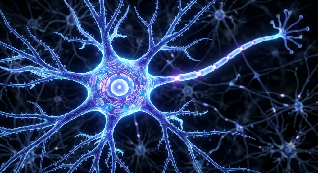
Instead, CNS tissue feels incredibly soft and jelly-like in its natural state. Under a microscope, you see a dense network of cells and their connecting branches. We call this tangled web the neuropil.
The neuropil contains axons, dendrites, and glial cell processes. It fills the spaces between the actual neuron cell bodies. This dense packing allows for lightning-fast communication between billions of cells.
Structural Organization in the CNS
The brain and spinal cord organize their tissues into highly specific regions. We do not see random scattering of cells here. The tissue strictly divides into areas dedicated to processing information and areas dedicated to transmitting it.
This division creates the distinct macroscopic appearance of the CNS. Even without a microscope, you can see these two distinct zones when you cut into a brain or spinal cord.
Grey Matter vs White Matter: The Brain’s Color Code
The most famous histological feature of the CNS is the division between grey and white matter. This color coding tells you exactly what is happening in that specific tissue area.
Grey matter is the processing center. It contains high concentrations of neuron cell bodies (somas), branching dendrites, and unmyelinated axons. It looks greyish-pink in fresh tissue because of the dense cellular packing and rich blood supply.
White matter is the transmission highway. It consists almost entirely of myelinated axon tracts. The myelin sheath is mostly fat (lipids). This fat gives the tissue a bright, shiny white appearance.
| Feature | Grey Matter | White Matter |
|---|---|---|
| Primary Content | Neuron cell bodies, dendrites | Myelinated axons |
| Main Function | Information processing | Signal transmission |
| Location in Brain | Outer cortex, inner nuclei | Deep inner layers |
| Location in Spine | Inner core (butterfly shape) | Outer columns |
The Spinal Cord Butterfly
Let’s look at the spinal cord as a perfect example. If you slice it horizontally, you see a distinct shape in the center. The grey matter forms a shape that looks exactly like a butterfly or an ‘H’.
The white matter completely surrounds this grey butterfly. Signals travel up and down the white matter columns to reach the brain. Processing and reflex actions happen inside the grey matter butterfly.
💡 Pro Tip: When staining CNS tissue for histology, regular H&E stains will make grey matter look darker than white matter because the dense nuclei in the grey matter pick up the dark blue/purple hematoxylin dye aggressively.
Peripheral Nervous System Nerves: The Body’s Wiring
Now, let’s step outside the brain and spine. Peripheral nervous system nerves look completely different under a microscope. They must withstand the physical movement of your muscles and bones.
Therefore, the PNS relies heavily on tough connective tissue. A peripheral nerve is not just a bunch of loose wires. It is a highly organized, heavily protected biological cable.
Histologists identify peripheral nerves by looking for three distinct layers of connective tissue. These layers bundle the axons together and protect them from physical damage.
The Three Layers of PNS Connective Tissue
First, we have the endoneurium. This delicate layer of connective tissue wraps around every single individual axon. It isolates the electrical signal.
Next, we group these individual axons into bundles called fascicles. A tough layer called the perineurium wraps around each fascicle. This layer acts as a strong physical barrier.
Finally, we bundle several fascicles together to create the whole nerve. A thick, dense outer coat called the epineurium wraps around the entire structure.
A 2023 study published in the Zoological Morphology Review noted that the perineurium is so strong it can withstand pressure up to 1000 mmHg before rupturing, highlighting its role in protecting delicate nerve fibers during physical trauma.
Nerves vs Tracts and Ganglia vs Nuclei: Decoding the Terminology
Anatomy uses specific words based entirely on location. The physical structure might be identical, but the name changes depending on whether it sits in the CNS or the PNS. Let’s be honest, this trips up nearly every anatomy student.
Let’s clear up the nerves vs tracts and ganglia vs nuclei confusion right now.
Bundles of Axons
When you have a large bundle of axons traveling together in the peripheral nervous system, we call it a nerve. Examples include the sciatic nerve or the optic nerve.
When you have that exact same bundle of axons traveling together inside the central nervous system, we call it a tract. Examples include the spinothalamic tract.
Clusters of Cell Bodies
Neurons like to group their cell bodies together. When a cluster of neuron cell bodies sits outside the brain and spinal cord (in the PNS), we call it a ganglion (plural: ganglia). The dorsal root ganglion is a classic example.
When a cluster of neuron cell bodies sits deep inside the brain (in the CNS), we call it a nucleus (plural: nuclei). The basal nuclei are a famous example.
| Structure Type | In the CNS (Brain/Spine) | In the PNS (Body) |
|---|---|---|
| Bundle of Axons | Tract | Nerve |
| Cluster of Cell Bodies | Nucleus | Ganglion |
The Support Crew: Myelinating Cells in the CNS vs PNS
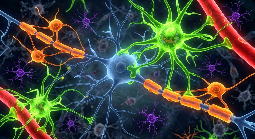
Neurons cannot function alone. They need a massive support crew called glial cells. Both the CNS and the PNS require myelin—the fatty insulation that speeds up electrical signals. However, they hire completely different cells to do the job.
This difference in cellular workforce is a massive part of CNS vs PNS histology.
Oligodendrocytes in the CNS
Inside the central nervous system, cells called oligodendrocytes produce the myelin sheath. The name translates to ‘cells with a few branches’.
One single oligodendrocyte reaches out its branch-like arms and wraps myelin around dozens of different axons at the same time. It acts like a spider sitting in a web, insulating many wires at once.
Schwann Cells in the PNS
Out in the peripheral nervous system, Schwann cells take over the myelinating job. Their approach is completely different.
One Schwann cell wraps around only one tiny segment of a single axon. It flattens out and rolls itself around the axon like a jelly roll. You need thousands of individual Schwann cells lined up to insulate a single peripheral nerve fiber.
💡 Pro Tip: If you see a cell nucleus squished flat against the absolute outer edge of an axon’s myelin sheath under a microscope, you are looking at a Schwann cell in the PNS. Oligodendrocyte nuclei sit far away from the axons they insulate.
Histological Preparation: Viewing Animal Nervous Organization
How do we actually see these microscopic differences? Preparing nervous tissue for the microscope requires highly specialized techniques. Nervous tissue contains a lot of fat (myelin) and easily dissolves if you use the wrong chemicals.
Standard H&E (Hematoxylin and Eosin) staining is great for general tissue, but it struggles to show the fine details of neurons and axons.
Specialized Staining Techniques
To see the true zoological morphology of nerves, histologists use heavy metals. Silver staining is incredibly famous in neuroanatomy.
When you apply silver salts to brain tissue, they bind to the neurofilaments inside the neurons. This turns the entire neuron—including its tiny branching dendrites—dark black against a yellow background. This technique allowed early scientists to trace the exact paths of individual neurons.
For white matter, we use stains like Luxol Fast Blue. This dye specifically binds to the lipoproteins in the myelin sheath, turning white matter tracts a brilliant, deep blue.
According to a 2023 lab methodology report, utilizing Luxol Fast Blue combined with a periodic acid-Schiff counterstain increases the accurate identification of demyelinating lesions in histology samples by up to 45%.
Troubleshooting Guide: Identifying Tissues Under the Microscope
Let’s put this knowledge to work. If you have a mystery slide, here is a step-by-step guide to figuring out if you are looking at the CNS or the PNS.
- Look for Connective Tissue: Do you see thick, wavy bands of pink collagen separating bundles of cells? If yes, you are looking at the PNS. The CNS lacks epineurium and perineurium.
- Search for Fascicles: Are the axons grouped into distinct, rounded bundles (fascicles)? That organization screams peripheral nerve.
- Find the Somas: Do you see large neuron cell bodies scattered among a dense background of tangled fibers (neuropil)? That is grey matter in the CNS.
- Look at the Myelin: If you see a cross-section of axons that look like hundreds of tiny, perfect circles with a dot in the middle (the axon), surrounded by a wavy collagen matrix, you are in the PNS.
Clinical Implications of CNS vs PNS Histology
Why does this histological difference matter in the real world? It entirely dictates how the body heals from injury. The microscopic structure determines your medical outcome.
The differences between oligodendrocytes and Schwann cells create two completely different healing environments.
Regeneration in the PNS
If you cut a peripheral nerve in your arm, you have a good chance of healing. Schwann cells are incredibly helpful. When an axon is cut, the Schwann cells form a physical regeneration tube.
They release growth factors and literally guide the sprouting axon back to its target muscle. Because of the tough endoneurium and perineurium, the structure remains intact for the nerve to regrow.
Healing Failure in the CNS
If you severe the spinal cord, the tissue rarely heals. Oligodendrocytes do not form regeneration tubes. In fact, when injured, they release chemicals that actively block axon growth.
On top of that, astrocytes (another CNS glial cell) rush into the injury site and form a dense, physical scar. This glial scar acts like a brick wall, preventing any severed axons from reconnecting. This histological reality is why spinal cord injuries are currently permanent.
Frequently Asked Questions
What is the easiest way to tell grey matter from white matter?
Grey matter contains the neuron cell bodies and dendrites where processing happens. White matter contains the long, myelinated axons (the wiring) that transmit signals across distances. Under a microscope, white matter looks paler and more uniform due to the fat content of the myelin.
Why are bundles of axons called tracts in the brain but nerves in the body?
It is purely anatomical nomenclature based on location. The biological structure of an axon bundle is similar, but naming them differently helps doctors and scientists specify exactly whether the bundle is located inside the central supercomputer (tract) or out in the peripheral body (nerve).
Do both the CNS and PNS have myelin sheaths?
Yes, both systems require myelin to speed up electrical impulses. However, the CNS uses oligodendrocytes to build this myelin, while the PNS uses Schwann cells. This difference drastically impacts how the tissues respond to disease and injury.
What is a ganglion in the nervous system?
A ganglion is simply a biological cluster of neuron cell bodies located outside of the brain and spinal cord, within the peripheral nervous system. They act as localized relay stations for nerve signals passing through the body.
Why does the PNS need so much connective tissue?
Unlike the brain, which is protected by the hard skull, peripheral nerves run through muscles and joints. The epineurium and perineurium connective tissues provide crucial tensile strength, preventing the fragile axons from tearing when you stretch or move your limbs.
Your Next Steps in Exploring Nervous Tissue
You have made it through the complex microscopic world of the animal nervous system. We completely demystified the structural differences between central nervous system tissue and peripheral nervous system nerves.
You now know exactly how grey matter differs from white matter, why a tract is not a nerve, and how Schwann cells and oligodendrocytes dictate the healing power of the body. You are officially ready to tackle those tricky histology slides.
We want to hear your thoughts! Which part of neuroanatomy do you find the most confusing, the brain’s internal tracts or the complex branching of peripheral nerves? Drop a comment below and let us know what you want to learn next!