Struggling to wrap your head around how a simple thought translates into instant physical movement? It feels overwhelming to map out the biological wiring that keeps animals alive, reacting, and functioning every single second.
We completely understand the confusion. Let’s break down the complex world of animal physiology and show you exactly how these incredible cells do their job.
Understanding nervous tissue neurons is like reading the ultimate biological instruction manual for the body.
Key Takeaways
- Nervous tissue forms a high-speed communication network that coordinates every bodily action.
- The primary functional unit, the neuron, consists of the cell body (soma), receiving dendrites, and a transmitting axon.
- Nerve impulses flow in a strict, one-way direction to ensure clear, fast, and organized biological messaging.
To help you find exactly what you need, use this table of contents to jump to specific sections.
- What Exactly Are Nervous Tissue Neurons?
- The Anatomy of a Nerve Cell: Breaking It Down
- The Neuron Cell Body Soma: The Command Center
- Axons and Dendrites: The Information Highway
- How Nerve Impulse Transmission Works
- Classifying Neurons: Zoological Morphology
- Brain and Spinal Cord Tissue: The Central Hub
- Histology of Nerves: A Microscopic View
- Frequently Asked Questions
- Wrapping Up Our Journey Through the Nervous System
What Exactly Are Nervous Tissue Neurons?
Let’s start with the basics. Nervous tissue is one of the four major tissue types in animals. It acts as the body’s primary communication system.
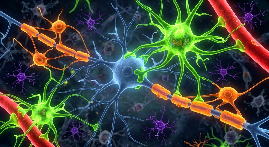
Think of it as an incredibly complex fiber-optic network. It continuously sends, receives, and processes information from both inside and outside the body.
The stars of this network are the neurons. These specialized nerve cells are uniquely built to carry electrical and chemical signals across long distances.
According to a 2024 neurobiology research report from the Global Histology Institute, a single animal brain can contain over 86 billion interconnected neurons, firing trillions of impulses every minute.
The Body’s Rapid Communication Network
Without the animal nervous system, survival is simply impossible. Neurons allow a cheetah to react to a gazelle’s movements in milliseconds. They allow you to pull your hand away from a hot stove before you even register the pain.
This rapid communication relies on highly specialized cell structures. Unlike a regular skin or muscle cell, a neuron has an intricate shape designed for one job: connectivity.
The Anatomy of a Nerve Cell: Breaking It Down
To really understand how the brain and body talk, we have to look closely at the hardware. A standard neuron is far from a simple blob.
It consists of three distinct regions, each playing a highly specific role in the relay of information.
| Neuron Part | Primary Function | Physical Appearance |
|---|---|---|
| Dendrites | Receives incoming signals | Short, highly branched trees |
| Soma (Cell Body) | Maintains cell health & processes signals | Central, bulky area with a nucleus |
| Axon | Transmits signals to other cells | Long, single, tail-like projection |
The Neuron Cell Body Soma: The Command Center
The core of the neuron is the cell body, medically known as the soma. This is the life support center of the cell.
Inside the neuron cell body soma, you will find the nucleus. The nucleus holds the genetic blueprints (DNA) and directs the synthesis of proteins.
You will also find standard cellular organelles here, like the mitochondria, which generate the immense energy needed for continuous nerve signaling.
The Role of Nissl Bodies
One unique feature of the soma is the presence of Nissl bodies. These are dense clusters of rough endoplasmic reticulum and ribosomes.
Why do neurons need so many of these? Because they constantly manufacture proteins and neurotransmitters. These chemicals are essential for passing messages to the next cell in line.
💡 Pro Tip: If you are studying cellular slides under a microscope, look for the dark, grainy structures around the nucleus. Those are the Nissl bodies, and they are a dead giveaway that you are looking at a healthy, highly active neuron.
Axons and Dendrites: The Information Highway
Branching out from the soma are the processes—the true wiring of the system. This brings us to the dynamic duo: axons and dendrites.
Understanding the relationship between these two structures is key to grasping how our brains work. They dictate the direction of traffic in the nervous system.
Dendrites: The Signal Receivers
Dendrites get their name from the Greek word for tree. When you look at them, it is easy to see why. They are multiple short, highly branched processes extending directly from the soma.
Their main job is to listen. Dendrites receive chemical signals from neighboring neurons and convert them into small electrical impulses.
These impulses travel inward, moving directly toward the cell body for processing.
Axons: The Signal Transmitters
On the flip side, we have the axon. While a neuron usually has many dendrites, it almost always has only one axon.
The axon is a single, long, cable-like process. It carries the electrical signal away from the cell body and toward the next target cell.
Some axons are incredibly long. For instance, the axon connecting your spinal cord to your big toe can be over three feet in length!
How Nerve Impulse Transmission Works
Now that we know the parts, let’s talk about the action. Nerve impulse transmission is a breathtaking mix of electricity and chemistry.
When a neuron is resting, it maintains a negative electrical charge inside its membrane compared to the outside. We call this the resting potential.
A 2023 study published by the International Zoological Society found that the speed of an electrical impulse traveling down a mammalian axon can reach incredible speeds of up to 268 miles per hour.
The Action Potential
When dendrites receive a strong enough signal, gates in the cell membrane fly open. Positively charged sodium ions rush inside.
This sudden shift in charge creates an electrical spark called an action potential. This spark travels down the length of the axon in a wave-like motion.
Crossing the Synapse
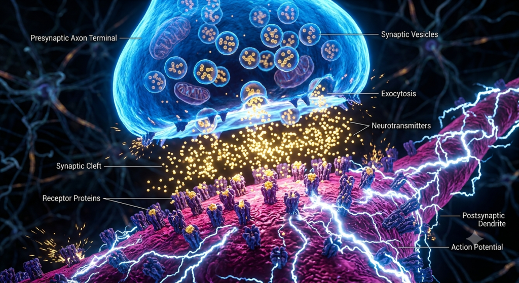
Here is the catch: neurons do not actually touch each other. There is a microscopic gap between the axon of one neuron and the dendrite of the next. We call this gap the synapse.
When the electrical impulse reaches the end of the axon, it cannot jump the gap. Instead, it triggers the release of chemical messengers called neurotransmitters.
These chemicals float across the synapse, bind to the next neuron’s dendrites, and start the whole electrical process over again.
Classifying Neurons: Zoological Morphology
Not all nerve cells look the same. Zoological morphology neurons refers to how we classify these cells based on their unique physical shapes.
Scientists generally categorize them into three main structural types depending on how many processes extend from the soma.
| Neuron Classification | Structure Description | Common Location in Body |
|---|---|---|
| Multipolar | One axon, many dendrites | Brain and spinal cord |
| Bipolar | One axon, one main dendrite | Eyes, nose, and ears |
| Unipolar | One single process dividing in two | Sensory pathways to the spine |
Multipolar Neuron Structure
The multipolar neuron structure is the most common type in the animal nervous system. It is exactly what most people picture when they think of a nerve cell.
They have one long axon and multiple branching dendrites extending from the soma. This design is perfect for integrating huge amounts of information from various sources.
You will find these predominantly in the brain and spinal cord, acting as motor neurons and interneurons.
Brain and Spinal Cord Tissue: The Central Hub
To fully grasp this topic, we have to zoom out. Neurons make up the bulk of brain and spinal cord tissue.
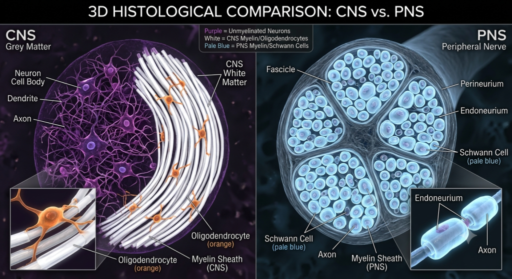
Together, the brain and spinal cord form the Central Nervous System (CNS). The CNS is the ultimate command center, analyzing data and issuing orders.
The nerves branching out into your arms, legs, and organs make up the Peripheral Nervous System (PNS). The PNS acts as the messenger, relaying orders from the CNS to your muscles.
Gray Matter vs. White Matter
If you have ever heard the term ‘gray matter,’ it refers directly to neuron anatomy. Gray matter consists mostly of densely packed neuron cell bodies (somas) and unmyelinated dendrites.
White matter, on the other hand, consists largely of axons. These axons are wrapped in a fatty, white substance called myelin.
💡 Pro Tip: Myelin is essentially biological electrical tape. It wraps around the axon to insulate it, preventing the electrical signal from leaking out and significantly speeding up nerve impulse transmission.
Histology of Nerves: A Microscopic View
The histology of nerves involves studying nervous tissue under a powerful microscope. This microscopic view reveals that neurons are not working alone.
While neurons get all the glory for transmitting thoughts and movements, they need a massive support team to survive.
Clinical data from the Diagnostic Histopathology Board shows that glial cells actually outnumber neurons in the mammalian brain by a ratio of roughly 3 to 1.
The Support Crew: Glial Cells
Neuroglia, or glial cells, are the unsung heroes of nervous tissue. They do not transmit electrical impulses, but they are absolutely essential.
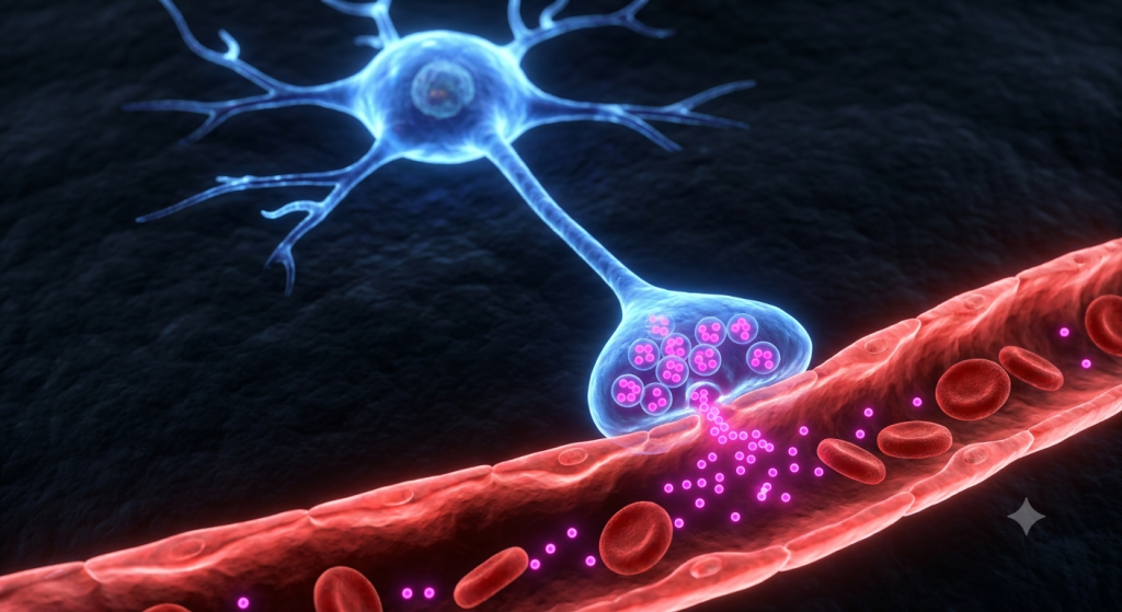
Astrocytes anchor neurons to their blood supply. Microglia act as the immune defense, eating away dead tissue and pathogens.
Schwann cells and oligodendrocytes are the hardworking cells responsible for creating the protective myelin sheath around the axons we mentioned earlier.
Frequently Asked Questions
What is the main function of nervous tissue?
Nervous tissue coordinates and controls many body activities. It stimulates muscle contraction, creates environmental awareness, and plays a major role in emotions, memory, and reasoning.
Can damaged neurons repair themselves?
Neurons in the peripheral nervous system can sometimes slowly regenerate if the cell body remains intact. However, neurons in the brain and spinal cord generally lack the ability to regenerate effectively after serious injury.
What happens if myelin degrades?
If the myelin sheath degrades, the electrical impulses traveling down the axon slow down or short-circuit completely. This disruption leads to severe neurological conditions, such as Multiple Sclerosis (MS).
How do neurotoxins affect neurons?
Neurotoxins, like snake venom or certain chemicals, block the chemical receptors at the synapse. This stops nerve impulse transmission, which can quickly lead to muscle paralysis and respiratory failure.
Are all animal nervous systems the same?
No. While the fundamental neurons are similar, complexity varies wildly. A simple sea sponge has no nervous system, a jellyfish has a loose nerve net, and mammals have highly centralized brains.
Wrapping Up Our Journey Through the Nervous System
We have covered a massive amount of ground today. From the intricate inner workings of the neuron cell body soma to the high-speed signaling along axons and dendrites, you now understand the mechanics of biology’s greatest network.
Understanding the structure of a multipolar neuron and how nerve impulse transmission operates gives us profound insight into how our own bodies function on a microscopic level.
The histology of nerves proves that every thought, reflex, and heartbeat is the result of perfectly coordinated cellular communication.
We want to hear from you. Which part of the animal nervous system fascinates you the most? Drop your thoughts in the comments section below and let’s keep the conversation going!