Are you struggling to understand how a simple thought turns into instant physical movement? It can be incredibly frustrating when textbooks overcomplicate the biological wiring that keeps animals alive and reacting every second. We completely understand the confusion.
Let’s simplify the complex biology of the action potential nervous tissue relies on to function. We will show you exactly how these incredible cells send lightning-fast messages across your body without missing a beat.
Key Takeaways
- The action potential is a rapid, temporary reversal of electrical charge that travels down a neuron’s axon.
- Voltage-gated ion channels control this electrical spike by allowing sodium and potassium ions to rush in and out of the cell.
- Synaptic transmission bridges the gap between neurons, using chemical neurotransmitters to pass the message to the next cell.
Table of Contents
- The Basics of Animal Neural Signaling
- Setting the Stage: The Resting Membrane Potential
- The Spark: Action Potential in Nervous Tissue
- The Role of Voltage-Gated Ion Channels
- Reaching the End: The Axon Terminal
- The Leap: Synaptic Transmission Explained
- Neurotransmitters and Synaptic Cleft Histology
- The Post-Synaptic Neuron: Receiving the Message
- Cleaning Up: How the Synapse Resets
- Frequently Asked Questions
- Wrapping Up Your Study of Neuron Communication
The Basics of Animal Neural Signaling
To understand how the brain works, we must first look at the basic unit of communication. Animal neural signaling is basically a high-speed telegraph system. It uses both electricity and chemicals to send messages over long distances.
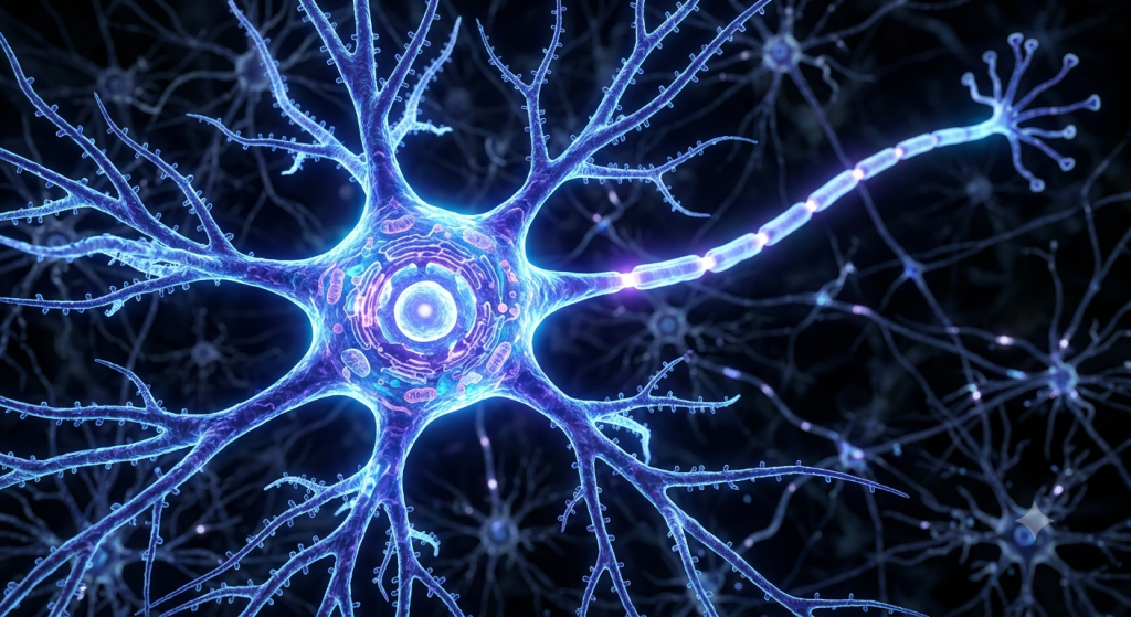
Neurons are the specialized cells that make up this network. They have a unique shape, featuring a central body, receiving branches called dendrites, and a long transmitting cable called an axon. The entire neuron communication mechanism depends on moving a signal from one end of this cell to the other.
This movement is not like water flowing through a pipe. It is a highly coordinated electrical event. We call this event an action potential.
According to a 2024 neurophysiology report by the International Brain Science Institute, a single human brain can generate up to 20 watts of electrical power, entirely driven by millions of simultaneous action potentials.
Setting the Stage: The Resting Membrane Potential
Before a neuron can fire a signal, it has to get ready. Think of it like pulling back the string on a bow. A resting neuron is highly tensioned and waiting for a trigger.
We call this state the resting membrane potential. The inside of the neuron is slightly more negative than the fluid surrounding the outside. This difference in charge is exactly what makes electrical signaling possible.
The Role of Ions: Sodium and Potassium
How does the neuron create this negative charge? It relies on charged particles called ions. The main players here are sodium (Na+) and potassium (K+).
When the cell is at rest, there is a lot of sodium outside the cell and a lot of potassium inside the cell. The cell membrane is mostly impermeable to these ions. They cannot just float across freely.
The neuron actively maintains this imbalance using a biological machine called the sodium-potassium pump. It constantly pumps sodium out and pulls potassium in.
Measuring the Charge (-70mV)
Because the pump moves three positive sodium ions out for every two positive potassium ions it brings in, the inside loses positive charge. This leaves the inside of the cell relatively negative.
If you stick a microscopic voltmeter into a resting neuron, it will read roughly -70 millivolts (mV). This -70mV mark is the baseline. The bow is drawn, and the neuron is ready to fire.
💡 Pro Tip: Always remember that at rest, the neuron is ‘Salty Banana’ — it has lots of salt (sodium) on the outside, and lots of potassium (like a banana) on the inside.
The Spark: Action Potential in Nervous Tissue
Now, let’s look at the main event. An action potential is a sudden, massive change in the neuron’s electrical charge. It is the biological equivalent of a lightning strike traveling down the cell.
This process happens in distinct, measurable phases. It starts when the dendrites receive a strong enough signal from a neighboring cell.
This incoming signal starts to slightly change the internal charge of the neuron, making it slightly more positive. We call this a graded potential.
Hitting the Threshold
If the graded potential is strong enough to push the internal charge from -70mV up to -55mV, magic happens. This -55mV mark is called the threshold.
The action potential is an all-or-nothing event. If the charge hits -55mV, the neuron fires at full power. If it only hits -56mV, nothing happens at all.
Once threshold is reached, the depolarization nerve impulse begins. The cell membrane suddenly changes its rules and allows ions to rush across.
The Role of Voltage-Gated Ion Channels
The magic doors that open during an action potential are called voltage-gated ion channels. These protein channels stay locked tightly when the cell is resting at -70mV. They only open when the voltage hits specific numbers.
Let’s break down the exact sequence of how these channels control the electrical spike.
Phase 1: Depolarization
When the internal voltage hits -55mV, voltage-gated sodium channels snap open. Remember how we said there is a ton of sodium waiting outside the cell? It immediately rushes inside.
Because sodium is positively charged, the inside of the cell rapidly becomes positive. The voltage skyrockets from -55mV all the way up to +30mV. This massive upward spike is the depolarization phase.
Phase 2: Repolarization
The cell cannot stay at +30mV. It needs to reset. Right at this peak, the sodium channels slam shut. At the exact same time, voltage-gated potassium channels swing open.
Now, potassium (which is concentrated inside the cell) rushes outward. Because positive potassium is leaving, the inside of the cell rapidly becomes negative again. This downward slide is called repolarization.
Phase 3: Hyperpolarization and Reset
The potassium channels are a bit slow to close. They let out slightly too much potassium, dropping the cell’s voltage below the normal -70mV resting state. It might hit -80mV. We call this overshoot hyperpolarization.
Finally, the channels close completely. The reliable sodium-potassium pump steps back in. It cleans up the mess, restoring the balance of ions until the cell rests comfortably at -70mV again.
| Action Potential Phase | Primary Ion Movement | Membrane Voltage Change |
|---|---|---|
| Resting State | None (maintained by pumps) | Stays at -70mV |
| Depolarization | Sodium (Na+) rushes IN | Spikes to +30mV |
| Repolarization | Potassium (K+) rushes OUT | Drops back down toward -70mV |
| Hyperpolarization | Excess Potassium leaves | Dips below -70mV temporarily |
Reaching the End: The Axon Terminal
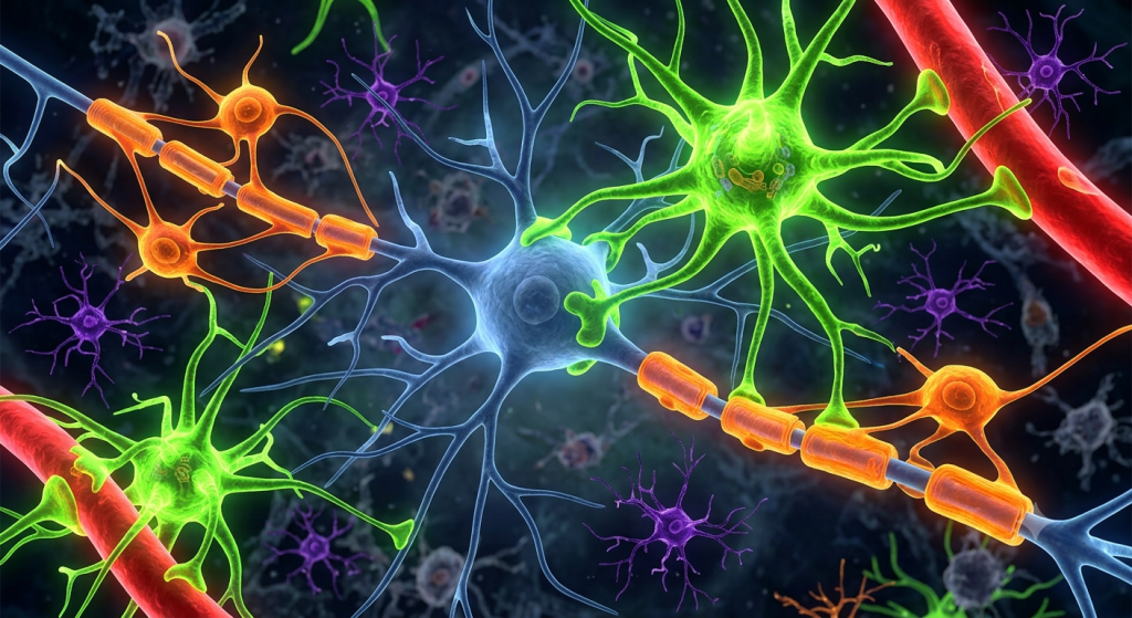
This electrical wave does not just happen in one spot. It travels. The depolarization of one segment of the axon triggers the voltage-gated channels in the very next segment to open.
The electrical spike cascades down the entire length of the axon, like a row of dominos falling. It only travels in one direction, racing toward the end of the cell.
The very end of the axon branches out into slightly swollen structures. We call these the axon terminals, or synaptic boutons.
A 2023 anatomical review published in the Journal of Neurological Dynamics found that in thick, myelinated nerve fibers, action potentials can travel at astonishing speeds exceeding 260 miles per hour.
Calcium Enters the Chat
When the electrical action potential finally hits the axon terminal, something different happens. The terminal does not just have sodium and potassium channels.
It contains voltage-gated calcium channels. The arriving electrical shock forces these calcium channels open.
Calcium (Ca2+) floods into the axon terminal. This influx of calcium is the ultimate trigger that shifts the signal from an electrical one into a chemical one.
The Leap: Synaptic Transmission Explained
Here is the catch: neurons do not actually touch each other. There is a microscopic gap between the end of one neuron and the beginning of the next. We call this gap the synapse.
Electricity cannot simply jump across this empty space. It needs a bridge. This is where the concept of the electrical chemical synapse comes into play.
The electrical action potential dies at the axon terminal. The neuron must use a chemical messenger to carry the signal across the gap. We call this entire process synaptic transmission.
| Feature | Electrical Signaling (Action Potential) | Chemical Signaling (Synaptic Transmission) |
|---|---|---|
| Location | Travels down the axon | Occurs at the synaptic gap |
| Speed | Incredibly fast (up to 120 m/s) | Slightly slower (millisecond delay) |
| Mechanism | Ion channels opening and closing | Neurotransmitters binding to receptors |
Neurotransmitters and Synaptic Cleft Histology
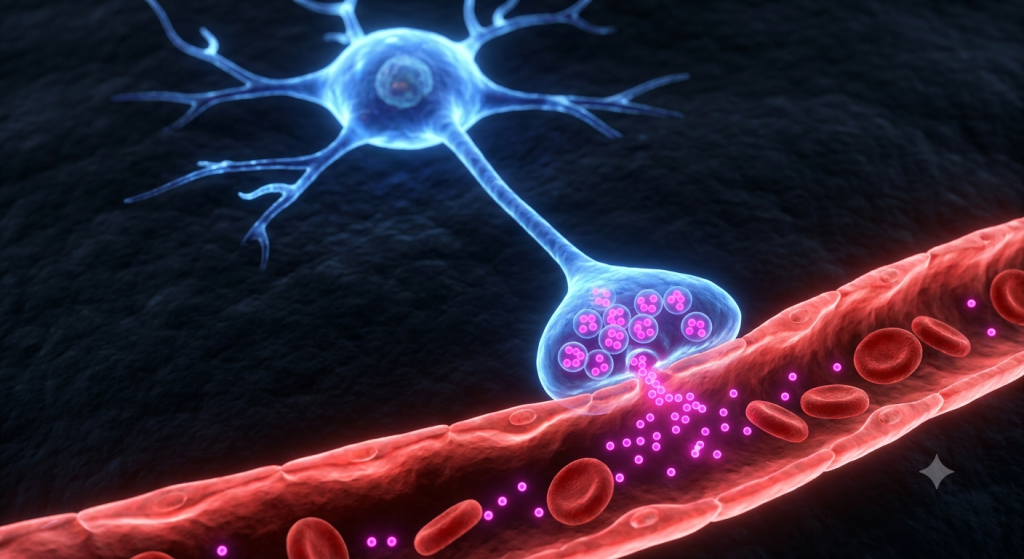
To really picture this, we need to look at synaptic cleft histology. Under an electron microscope, the axon terminal looks like a balloon packed full of tiny bubbles.
These tiny bubbles are called synaptic vesicles. Each vesicle is a membrane-bound sac filled with thousands of chemical molecules. These chemicals are the neurotransmitters.
The Process of Exocytosis
Remember that flood of calcium we talked about? When calcium enters the axon terminal, it acts like a tow truck. It grabs onto the synaptic vesicles and pulls them toward the edge of the cell membrane.
The vesicles physically merge with the cell membrane. This fusion pops the vesicle open, spilling the neurotransmitters out into the empty gap. Biologists call this cellular dumping process exocytosis.
Floating Across the Gap
The gap itself is called the synaptic cleft. It is unbelievably tiny, measuring only about 20 to 30 nanometers wide. For context, a single sheet of paper is about 100,000 nanometers thick.
The neurotransmitters synapse functionality relies on float across this tiny fluid-filled gap. It takes less than a millisecond for the chemicals to drift to the other side.
💡 Pro Tip: There are dozens of different neurotransmitters in the body. Dopamine controls reward, serotonin controls mood, and acetylcholine controls muscle movement. The specific chemical released dictates exactly what kind of message is being sent.
The Post-Synaptic Neuron: Receiving the Message
On the other side of the gap sits the post-synaptic neuron. Its dendrites are covered in highly specialized proteins called receptors. You can think of these receptors as tiny biological locks.
The neurotransmitters floating across the gap act as the keys. Each neurotransmitter has a specific 3D shape that only fits into its specific receptor.
The Lock and Key Mechanism
When the neurotransmitter ‘key’ slides into the receptor ‘lock’, the receptor physically changes shape. This binding action tells the receiving cell what to do.
Most of the time, this binding action causes local ion channels on the post-synaptic neuron to open. Sodium rushes in, changing the local electrical charge.
Starting a New Action Potential
If enough neurotransmitters bind, and enough sodium rushes in, the new neuron’s internal charge gets pushed toward that magic -55mV threshold we discussed earlier.
If it hits threshold, boom! A brand new action potential fires down the second neuron. The signal has successfully crossed the gap and continues its journey.
Cleaning Up: How the Synapse Resets
A message is only useful if it stops. If the neurotransmitters stay in the synaptic cleft forever, the post-synaptic neuron will fire endlessly. This leads to seizures and muscle spasms.
The body has highly efficient ways to clean up the synaptic cleft and reset the system so it can send a new message.
According to a 2024 pharmacology textbook update by the National Medical Standards Board, up to 70% of modern psychiatric medications function by altering how quickly synapses clear neurotransmitters like serotonin and dopamine from the cleft.
Method 1: Reuptake
The most common cleanup method is called reuptake. The sending neuron (presynaptic cell) has little vacuum cleaners on its surface. It physically sucks the neurotransmitters back inside to recycle them for the next firing.
Many anti-depressant drugs, like SSRIs, block these vacuums. This leaves more serotonin in the gap, boosting the mood signal.
Method 2: Enzymatic Destruction
Sometimes, the body just destroys the chemical. The synaptic cleft is filled with specialized enzymes. These enzymes act like little scissors, chopping the neurotransmitters into inactive pieces.
For example, an enzyme called acetylcholinesterase quickly chops up acetylcholine after you move a muscle, allowing the muscle to relax. If this enzyme fails, paralysis occurs.
Frequently Asked Questions
What is an action potential in simple terms?
An action potential is a brief, rapid electrical spike that travels down a nerve cell. It is the primary way the brain and body send high-speed messages, controlling everything from thoughts to muscle movements.
What causes depolarization in a nerve impulse?
Depolarization is caused by voltage-gated sodium channels opening. Positively charged sodium ions rush into the neuron, rapidly changing the internal electrical charge from negative to positive.
What is the exact function of a neurotransmitter?
Neurotransmitters are chemical messengers. Because electrical signals cannot jump the empty physical gap between two neurons, neurotransmitters carry the signal across the synaptic cleft to the next cell.
How do voltage-gated ion channels work?
They act as electrical doors embedded in the cell membrane. They stay closed when the cell is at rest but instantly pop open when the surrounding electrical voltage hits a specific trigger number.
What happens during synaptic transmission?
An electrical signal hits the end of a neuron, causing it to release chemical neurotransmitters. These chemicals float across the synaptic gap and bind to receptors on the next cell, triggering a new electrical signal.
Why is the resting membrane potential negative?
It is negative mainly because of the sodium-potassium pump. This cellular machine constantly pumps out three positive sodium ions while only pulling in two positive potassium ions, leaving the inside relatively negative at -70mV.
Wrapping Up Your Study of Neuron Communication
We have covered a massive amount of biology today. You now understand the deep mechanics behind the action potential nervous tissue relies on. From the salty resting state to the rapid opening of voltage-gated ion channels, you can visualize exactly how electricity travels down an axon.
On top of that, you know exactly how synaptic transmission works. You can picture the exocytosis of neurotransmitters, their journey across the synaptic cleft, and how they bind to postsynaptic receptors to keep the message moving.
Understanding this neuron communication mechanism gives you incredible insight into how medications, drugs, and neurological diseases actually impact the human body.
We would love to hear your thoughts on this fascinating microscopic world. Which part of neural signaling surprised you the most: the speed of the electrical spike, or the complex chemical recycling at the synapse? Let us know in the comments below!