Do you struggle to understand how the brain actually maintains itself against toxins and damage? It can be incredibly frustrating when biology textbooks only talk about flashy neurons, completely ignoring the vital support structures that keep them alive. We are here to fix that by revealing the true power of neuroglia glial cells.
Without these microscopic bodyguards, our nervous system would collapse in minutes. Let’s break down the hidden world of nervous tissue support cells.
Key Takeaways
- Neuroglia outnumber neurons significantly and provide structural, metabolic, and immune support to the entire nervous system.
- Astrocytes maintain the blood-brain barrier, filtering nutrients and blocking deadly toxins from reaching your vulnerable brain cells.
- Schwann cells and oligodendrocytes create insulating myelin sheaths, which drastically speed up electrical signals across your body.
To help you find exactly what you need quickly, use this table of contents to jump directly to specific topics.
- The Hidden Majority: What Are Neuroglia Glial Cells?
- Astrocytes and the Blood Brain Barrier
- Schwann Cells: The Myelin Makers of the PNS
- Oligodendrocytes: Central Nervous System Insulation
- Microglia: The Immune Defense of the Brain
- Other Vital Support Cells in Nervous Tissue
- Why Neuron Protection and Nourishment Matters
- A Step-by-Step Look at Glial Support During Neural Firing
- Animal Brain Histology: Viewing Glia Under the Microscope
- Frequently Asked Questions
- Wrapping Up Our Microscopic Journey
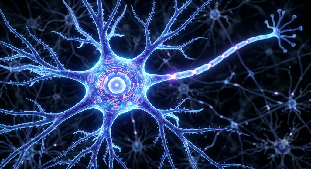
The Hidden Majority: What Are Neuroglia Glial Cells?
When people think of the brain, they picture neurons firing electricity. We celebrate neurons as the stars of the show. However, neurons are actually high-maintenance divas.
They demand constant food, extreme protection, and a perfectly balanced chemical environment. If they do not get these things, they die rapidly.
This is exactly where neuroglia glial cells step in. The word ‘glia’ literally translates to ‘glue’ in Greek. Early anatomists thought these cells just held the brain together.
Today, we know they do so much more than act as biological paste. They manage the entire background operation of your thoughts, movements, and memories.
According to a 2024 cellular biology report from the Global Neuroscience Institute, neuroglia make up roughly 50 to 90 percent of all cells in the human brain, completely dwarfing the neuron population.
Glial Cells vs. Neurons: The Big Difference
So, how do these two cell types differ? The most important distinction lies in electricity. Neurons generate and conduct action potentials.
Neuroglia do not generate electrical impulses. They cannot send a message to your muscles to move your arm.
Instead, they set the stage. They clean up chemical spills, provide physical scaffolding, and wrap the biological wires to prevent short circuits.
💡 Pro Tip: If you are studying for a biology exam, remember this simple rule: Neurons communicate, while glia support and regulate. Keeping this core difference in mind helps you answer most functional anatomy questions.
Astrocytes and the Blood Brain Barrier
Let’s look at the most abundant glial cell in the central nervous system (CNS). Astrocytes get their name from their striking, star-like appearance.
They have dozens of long, branching arms that stretch out in every direction. These arms grab onto neurons on one side and blood vessels on the other.
This physical connection allows astrocytes to control the flow of life-saving materials.
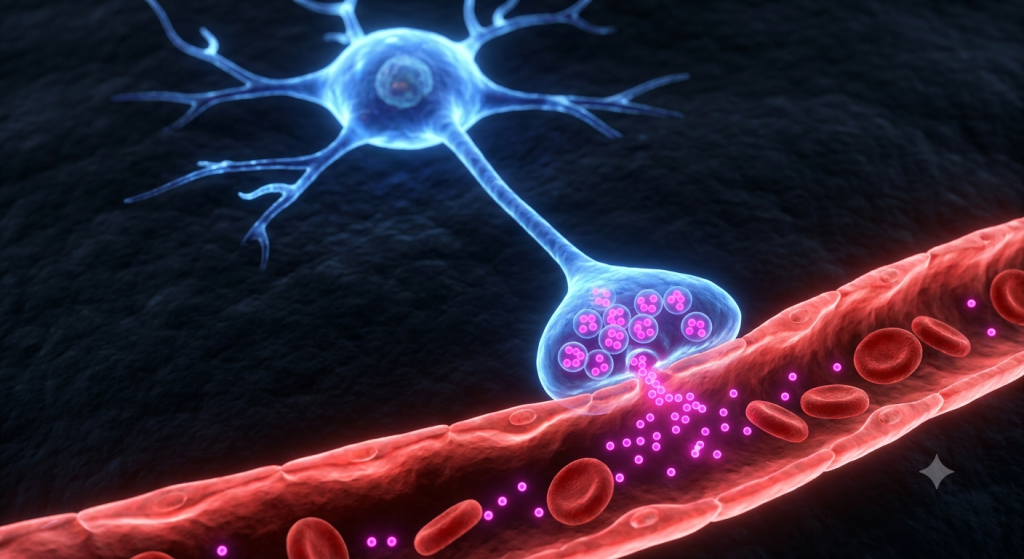
Building the Blood-Brain Barrier
The blood-brain barrier is a highly selective filter. It separates the circulating blood from the brain’s extracellular fluid.
Astrocytes wrap their tiny ‘feet’ around the microscopic blood capillaries in the brain. They tell the capillary walls to form tight junctions.
This tight seal prevents bacteria, viruses, and large chemical molecules from leaking out of the blood and damaging the delicate neurons.
Metabolic Support and Nutrient Filtering
Astrocytes do not just block the bad stuff. They actively pull the good stuff out of your blood.
They extract glucose from the capillaries, convert it into lactate, and feed it directly to the hungry neurons. This process ensures neurons have the energy they need to fire continuously.
They also mop up extra potassium ions that leak out when neurons fire. If potassium builds up too much, neurons lose their ability to send signals.
Schwann Cells: The Myelin Makers of the PNS
Now, let’s step outside the brain and spinal cord. The peripheral nervous system (PNS) includes all the nerves running to your arms, legs, and organs.
Here, a different type of glial cell takes over. Schwann cells are the primary support staff for peripheral nerves.
Their main job is creating the myelin sheath. Myelin is a fatty, white substance that acts like rubber insulation around a copper electrical wire.
A 2023 anatomical survey published in the Journal of Peripheral Neurobiology found that a single Schwann cell can wrap its membrane around an axon up to 100 times to create a fully mature myelin sheath.
How Schwann Cells Wrap Axons
A Schwann cell finds a naked axon and flattens itself out. It then begins to roll around the axon like a bandage wrapping a sprained ankle.
It squeezes all its internal cytoplasm to the outer edges. This leaves behind tightly packed layers of fat and protein directly on the axon.
This insulation prevents the electrical signal from leaking out into the surrounding tissue. It keeps the signal strong and focused.
Regeneration and Repair in Peripheral Nerves
Schwann cells have a superpower that central nervous system glia lack. They actively help damaged nerves heal.
If you cut your finger and sever a nerve, the Schwann cells in that area form a regeneration tube. They guide the broken axon back to its original target.
They secrete growth factors that encourage the neuron to sprout new branches. This is why you can regain feeling in a deep cut after a few months.
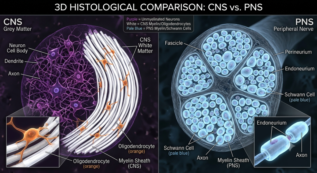
Oligodendrocytes: Central Nervous System Insulation
Back inside the brain and spinal cord, we find oligodendrocytes. The name literally means ‘cells with a few branches.’
These cells serve the exact same purpose as Schwann cells. They build myelin sheaths to insulate axons.
However, their method of building this insulation looks completely different.
The Multi-Tasking Wrappers
A Schwann cell dedicates its entire life to wrapping just one tiny section of one single axon. Oligodendrocytes are much more efficient.
One oligodendrocyte sits in the middle of a group of neurons. It sends out multiple flat extensions, like an octopus reaching out with its tentacles.
Each tentacle grabs a different axon and wraps a segment of myelin around it. A single oligodendrocyte can insulate up to 50 different axons at once.
| Feature | Schwann Cells | Oligodendrocytes |
|---|---|---|
| Location in Body | Peripheral Nervous System (PNS) | Central Nervous System (CNS) |
| Myelination Ratio | 1 cell insulates 1 axon segment | 1 cell insulates up to 50 axons |
| Regeneration Ability | Actively guides nerve regrowth | Inhibits nerve regrowth after injury |
Speeding Up Nerve Impulses
The myelin sheath created by these cells is not continuous. It has tiny gaps called the Nodes of Ranvier.
The electrical signal does not travel smoothly down the wire. Instead, it jumps rapidly from gap to gap.
We call this jumping process saltatory conduction. It makes nerve impulses travel up to 100 times faster than they would on an uninsulated axon.
Microglia: The Immune Defense of the Brain
The brain is a restricted VIP club. Standard immune cells, like white blood cells, are not allowed past the blood-brain barrier.
So, how does the brain fight off infections? It relies on a special type of glial cell called microglia.
Microglia are tiny, spidery cells that constantly patrol the brain and spinal cord, looking for trouble.
Phagocytosis and Cleaning Up Debris
Microglia act as the brain’s personal garbage collectors and security guards. When they find a dead cell, a tangle of proteins, or a trespassing bacteria, they transform.
They retract their spidery arms, become round, and physically swallow the threat. We call this cell-eating process phagocytosis.
They digest the harmful material using powerful internal enzymes, keeping the brain tissue pristine and healthy.
💡 Pro Tip: Researchers now believe that hyperactive microglia might contribute to neurodegenerative diseases like Alzheimer’s. If they become too aggressive, they start eating healthy synapses instead of just cleaning up debris.
Other Vital Support Cells in Nervous Tissue
While astrocytes, Schwann cells, oligodendrocytes, and microglia get most of the attention, the nervous tissue support system relies on a few other key players.
These specialized cells handle highly specific tasks that keep the environment perfectly balanced.
Ependymal Cells and Cerebrospinal Fluid
Deep inside the brain, there are hollow cavities called ventricles. Ependymal cells line the walls of these cavities.
These cells have tiny, hair-like structures called cilia on their surface. They constantly beat these cilia back and forth.
This movement helps circulate cerebrospinal fluid (CSF) throughout the central nervous system. Ependymal cells also play a role in actually producing this vital, shock-absorbing fluid.
Satellite Cells in the Peripheral Ganglia
Satellite cells are small, flat cells found in the peripheral nervous system. They wrap around the large cell bodies of sensory neurons located in clusters called ganglia.
They act very much like astrocytes do in the brain. They regulate the chemical environment, supplying nutrients and absorbing heavy metals to protect the vulnerable cell body.
| Glial Cell Type | Primary Function | Location |
|---|---|---|
| Astrocytes | Nutrient supply & Blood-Brain Barrier | CNS |
| Microglia | Immune defense & phagocytosis | CNS |
| Ependymal Cells | Circulate cerebrospinal fluid | CNS |
| Satellite Cells | Regulate chemical environment | PNS |
Why Neuron Protection and Nourishment Matters
Understanding these support cells reveals a massive truth about biology. Neurons are incredibly fragile.
Without constant neuron protection and nourishment, our bodies would fail almost instantly. The entire system relies on a delicate balance of chemicals and physical barriers.
A recent 2024 pathological study by the National Glial Research Board noted that a failure in astrocyte metabolic support can lead to permanent neuronal death in less than 5 minutes during a stroke.
The Consequences of Glial Failure
What happens when the glia stop working? The results are usually devastating neurological diseases.
Take Multiple Sclerosis (MS), for example. In MS, the body’s own immune system attacks the oligodendrocytes in the brain and spinal cord.
The myelin sheath degrades, exposing the bare axons. The electrical signals slow down, short-circuit, or stop completely, leading to muscle weakness, blindness, and paralysis.
How Glia Keep You Thinking Clearly
On a more positive note, healthy glia make you smarter. Astrocytes literally shape the way neurons connect to each other.
When you learn a new skill, astrocytes help strengthen the synapses between the neurons involved. They release chemicals that make the connection faster and more permanent.
Without this active glial participation, you would struggle to form new memories or learn new physical tasks.
A Step-by-Step Look at Glial Support During Neural Firing
Let’s look at a real-world scenario. Imagine you decide to kick a soccer ball. Here is how the glia support that single, rapid action.
This process highlights just how integrated these support cells are with the neurons.
Step 1: Pre-Firing Environment Check
Before the motor neuron even fires, astrocytes are hard at work. They are pulling glucose from the blood and delivering it to the neuron.
Satellite cells in the peripheral nervous system ensure the chemical balance around the neuron’s cell body is perfectly primed for action.
Step 2: Insulating the Signal
Your brain sends the command. The electrical action potential races down the axon toward your leg muscles.
Oligodendrocytes in your spinal cord and Schwann cells in your leg ensure the signal does not leak. The impulse jumps between the Nodes of Ranvier, reaching your muscle in milliseconds.
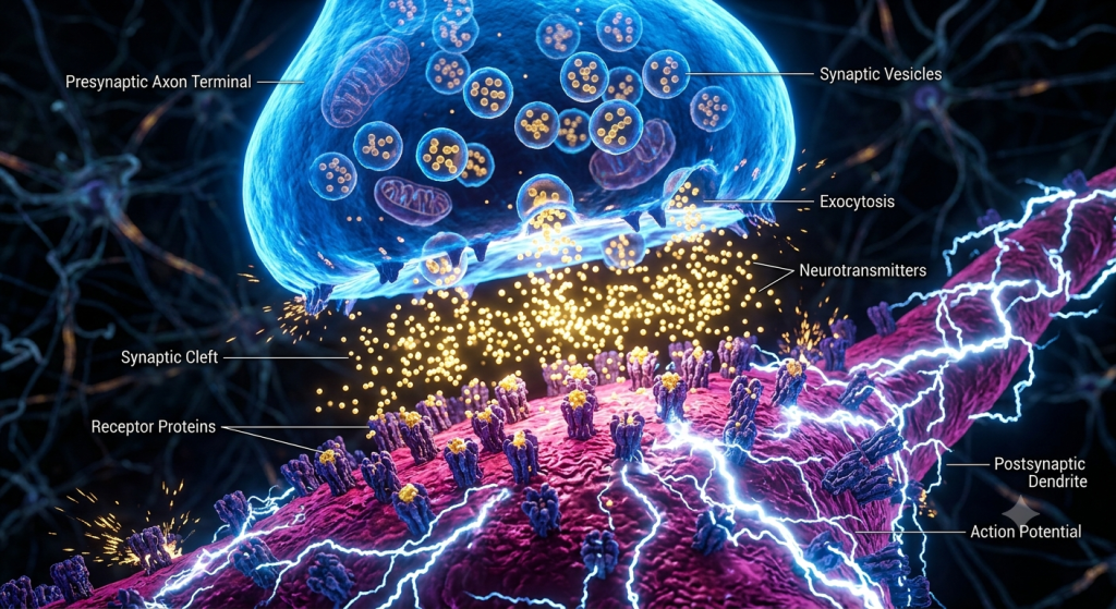
Step 3: The Cleanup Crew Arrives
After the neuron fires, it releases a flood of potassium ions and neurotransmitters into the surrounding space.
Astrocytes rush in. They vacuum up the excess potassium so the neuron can reset. They also absorb leftover neurotransmitters, repackage them, and hand them back to the neuron to use for the next kick.
Animal Brain Histology: Viewing Glia Under the Microscope
Historically, neuroglia were incredibly difficult to study. They are practically invisible under standard microscopes.
Animal brain histology changed dramatically when scientists developed special silver staining techniques in the late 19th century.
These stains finally allowed researchers to see the complex, branching structures of astrocytes and microglia.
Identifying Different Glia
Today, histologists use advanced fluorescent dyes and antibodies to tell the cells apart.
If a scientist wants to see astrocytes, they use a stain that attaches to a specific protein called GFAP. Suddenly, thousands of bright, star-shaped cells light up on the slide.
Microglia look completely different. Under a stain, they appear as small, dark, hairy little spots scattered randomly between the large neuron bodies.
💡 Pro Tip: When looking at histological slides of white matter, you are mostly seeing the lipid (fat) rich myelin sheaths created by oligodendrocytes. The fat does not absorb normal stains well, which is why the tissue looks white and pale to the naked eye.
Frequently Asked Questions
What is the main function of neuroglia?
Neuroglia provide essential physical structure, metabolic nourishment, immune defense, and electrical insulation for neurons. They ensure the nervous system operates smoothly by maintaining a healthy, balanced cellular environment.
Do glial cells transmit nerve impulses?
No. Unlike neurons, glial cells do not generate or transmit electrical action potentials. Their role is strictly supportive, regulating the environment so neurons can fire correctly.
Can neuroglia regenerate after an injury?
Yes. Glial cells retain the ability to divide and multiply throughout your life. In fact, when brain tissue is damaged, astrocytes rapidly multiply to form a glial scar to protect the area.
What causes a brain tumor?
Because mature neurons generally cannot divide, most brain tumors do not come from neurons. They primarily originate from mutated glial cells that multiply uncontrollably. These are called gliomas.
Why is the myelin sheath so important?
The myelin sheath insulates the axon, preventing electrical signals from leaking. This allows nerve impulses to travel incredibly fast and efficiently across long distances in the body.
What is the difference between Schwann cells and oligodendrocytes?
Schwann cells produce myelin in the peripheral nervous system (body), wrapping one axon segment per cell. Oligodendrocytes produce myelin in the central nervous system (brain/spine), wrapping multiple axons at once.
Wrapping Up Our Microscopic Journey
We have explored the hidden scaffolding of the brain. From the blood-brain barrier managed by astrocytes to the rapid immune responses of microglia, it is clear that neurons cannot survive alone.
Understanding the role of neuroglia gives us a complete picture of animal brain histology. It shows us that true brain power relies on teamwork, insulation, and constant microscopic maintenance.
The next time you quickly catch a falling object, do not just thank your neurons. Thank the billions of Schwann cells and oligodendrocytes that insulated the wires to make that reflex possible.
We would love to hear your thoughts on this complex microscopic world. Which of the glial cells do you think has the most fascinating job in the body? Let us know in the comments below!