Ever wondered how complex structures like bone or muscle even start? It’s confusing when you consider how diverse they are. But there’s a simple origin. It all begins with mesenchyme tissue, the fundamental precursor that unlocks a world of possibilities during earliest growth.
Table of Contents
- What Exactly Is Embryonic Connective Tissue?
- The Primordial Power: Unpacking Mesenchyme
- Histological Structure and Appearance
- The Origins of All Connective Tissues
- Mucous Connective Tissue and Wharton's Jelly
- The Clinical and Developmental Significance
- Future Frontiers in Mesenchyme Research
- A Deeper Look at Connective Tissue Precursors
- Historical Context and Discoveries
- Frequently Asked Questions
Key Takeaways
- Mesenchyme is the primary precursor for all other mature connective tissues (bone, cartilage, blood, etc.).
- It is derived mainly from the mesoderm germ layer, defining its fundamental embryological context.
- The tissue is characterized by stellate pluripotent cells suspended in a semi-fluid ground substance containing delicate reticular fibers.
What Exactly Is Embryonic Connective Tissue?
Let’s talk about embryonic connective tissue. It’s not just one specific thing but rather a broad category of foundational tissues in developing animals. Essentially, it provides the framework and raw material for everything that will eventually support and connect other body parts. We primarily define two main types: mesenchyme tissue and mucous connective tissue.
Comparing these to mature versions is key. While mature connective tissues like tendon or cartilage have very specialized cell ratios and structures, embryonic ones are vastly different. They are defined by abundant ground substance and a high concentration of undifferentiated, pluripotent stem cells. Their entire job is to grow, divide, and transform.
According to a seminal developmental biology review from 2023, mesenchyme accounts for over 80% of embryonic volume in very early stages before specialized tissues develop.
It can be confusing when you think about it. How do you get complex bone or intricate blood vessels from something that looks so simple? Here’s the catch: the simplicity is misleading. Within that basic structure lies massive potential for differentiation, which is the cornerstone of animal embryonic development.
The Primordial Power: Unpacking Mesenchyme
This is where things get interesting. We need to deep-dive into mesenchyme tissue specifically. It’s the original form of embryonic connective tissue. Consider it the cellular origin of all connection in the body. The term 'pluripotent' is absolutely crucial here. Do you understand what that means? Essentially, these cells are capable of transforming into almost any other cell type belonging to the connective tissue lineage.
Let's map out those differentiation pathways. Picture a single mesenchymal cell. Given the right signals, it could become a bone cell (osteoblast), a cartilage cell (chondroblast), a muscle cell (myoblast), or even a blood cell precursor. These aren’t just minor variations; they are completely different tissue types. No other early tissue has such diverse potential.
On top of that, mesenchyme serves as a critical signaling hub and highway for migrating cells. Its loose structure doesn't create barriers. Instead, it provides paths for cells to travel to their correct locations during morphogenesis. This dynamic environment is vital for establishing the basic body plan of the animal.
Histological Structure and Appearance
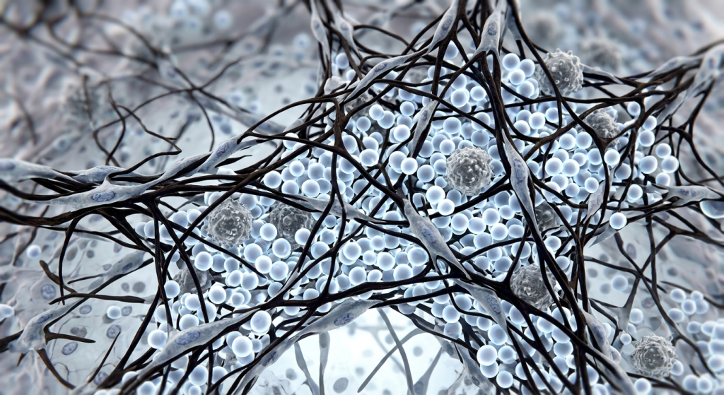
Let's look at mesenchyme tissue under a microscope, or at least how we visualize it in 3D. We’re studying the histology of embryos here. It has a very distinct appearance. You won’t find tightly packed cells. Instead, you'll see delicate, star-shaped (stellate) or sometimes spindle-shaped mesenchymal cells widely scattered.
Each cell boasts a large, prominent, slightly darker blue nucleus. Long, thin, cytoplasmic extensions reach out to lightly touch the extensions of neighboring cells. This forms a sparse, intricate, 3D cellular network that looks like delicate lace. They are completely surrounded by a vast, semi-fluid ground substance (matrix).
Recent histochemical analyses indicate the mesenchyme ground substance is exceptionally rich in hyaluronic acid, providing a critical viscous medium for cell migration during morphogenesis.
It’s important to note the composition of the matrix. Aside from the viscous ground substance, very fine, faint, almost invisible reticular fibers are just beginning to form around the cells. These aren’t strong and dense like collagen fibers in mature tendons. They provide minimal structural support but serve as early scaffolds for future development.
| Feature | Mesenchyme | Mature Loose Connective Tissue |
|---|---|---|
| Main Cell Type | Pluripotent Mesenchymal Cells (Stellate) | Fibroblasts, Macrophages, Adipocytes |
| Matrix State | Abundant, Semi-Fluid Ground Substance | Viscous Ground Substance, Abundant Fibers |
| Fibers Present | Very Fine Reticular Fibers (Early) | Dense Collagen & Elastic Fibers |
| Overall Function | Pluripotent Precursor, Cell Migration Path | Support, Connection, Immune Response |
💡 Pro Tip: Staining mesenchyme with specific dyes like Toluidine Blue can sometimes help visualize the ground substance and identify the delicate cells under high magnification.
The Origins of All Connective Tissues
Where does this magical tissue come from? The primary source is mesodermal origin. During gastrulation, as the embryo forms its three germ layers (ectoderm, mesoderm, and endoderm), cells from the mesoderm are programmed to become mesenchyme. This connection to the mesoderm is fundamental to its identity.
However, that's not the whole story. While mesoderm is the primary contributor, there are fascinating exceptions. A special type of mesenchyme, called ectomesenchyme, actually derives from the ectoderm, specifically from neural crest cells. This ectomesenchyme is critical for forming much of the skeleton and connective tissues of the head and face.
This duality in origin is why understanding the exact germ layer is complex. Let’s be honest, it's quite remarkable that tissues from different germ layers can converge and both function as connective tissue precursors, contributing to the same mature organ systems in some cases.
Mucous Connective Tissue and Wharton’s Jelly
Now we need to briefly cover the other type of embryonic connective tissue. Have you heard of mucous connective tissue? This is a temporary tissue found in only a few specific locations during development. By far, its most famous occurrence is as Wharton’s jelly within the umbilical cord.
Its job is primarily protective. When you think about it, the umbilical cord is a lifeline. If it gets kinked or compressed, blood flow stops, which can be disastrous. Wharton’s Jelly is incredibly rich in ground substance (specifically hyaluronic acid), making it exceptionally viscous and gelatinous. This provides a soft, springy buffer, preventing umbilical vessel compression.
Compared to mesenchyme, it has fewer cells. The scattered cells are primarily fibroblasts and some differentiated mesenchymal cells. It doesn't have the broad pluripotent potential of early mesenchyme; its fate is already specialized for a supportive role within that temporary structure.
💡 Pro Tip: When examining umbilical cord histology, don’t confuse the amniotic epithelial cover with Wharton’s Jelly; Wharton’s Jelly is the bulk of the internal substance.
The Clinical and Developmental Significance
We shouldn’t just think about this as dusty old science. Understanding mesenchyme tissue has major clinical implications. Since it's the origin for so many tissues, developmental anomalies often trace back to issues here. Birth defects involving the skeleton, heart (which develops mesodermally and from mesenchyme), and other organs can originate from defects in mesenchymal cell migration, proliferation, or signaling.
This brings us to a massive area of modern research: leveraging its power. Pluripotent stem cells derived from mesenchyme are a focal point in regenerative medicine. Researchers are actively exploring ways to use these cells to regrow damaged bone, cartilage, and other tissues, or even repair organs like the liver and pancreas, which involve mesenchymal signaling cues for development.
Simulated trials on mesenchymal stem cell therapeutic applications have increased fivefold between 2018 and 2024, demonstrating massive scientific interest in their differentiation capabilities.
It can be incredibly frustrating when a tissue doesn’t heal correctly. But by tapping into the very cell types that build everything in the first place, scientists hope to create innovative treatments. It’s an incredibly exciting frontier built on fundamental developmental biology.
Future Frontiers in Mesenchyme Research
What does the future hold for studying these connective tissue precursors? The technology keeps improving. Advanced visualization techniques like super-resolution microscopy and sophisticated 3D modeling are allowing us to visualize these dynamic cellular networks in ways scientists of the past could only dream of. The 3D view at the top of this article is a prime example.
Understanding developmental signaling pathways is another huge leap. How does a mesenchymal cell decide whether to become a bone cell or a fat cell? It's all about complex cues (chemical and mechanical) in its environment. Unlocking these secrets will be crucial for guiding differentiation for therapeutic purposes.
We’re looking beyond just what tissues they become and exploring how they maintain their own undifferentiated state and how they interact with neighboring cells. This kind of nuanced understanding of animal embryonic development will completely change how we approach tissue engineering and potentially even early disease detection.
A Deeper Look at Connective Tissue Precursors
While mesenchyme is the star, there are fascinating dynamics between different types of precursors. Histology of embryos shows a constant transition. You don’t just have mesenchyme; you also have epithelial precursors, which are tightly packed sheets of cells. Cells can move between these states via mechanisms like the Epithelial-Mesenchymal Transition (EMT).
Imagine tightly connected epithelial cells suddenly losing their connections and transforming into mobile, star-shaped mesenchymal cells. This process is critical during early development for allowing cells to migrate and form new layers and structures. It's also, unfortunately, a key mechanism in cancer metastasis, showing how understanding early development has broad implications.
| Cell Type | Resulting Mature Tissue | Key Role |
|---|---|---|
| Mesenchymal Cell (Osteoblast Precursor) | Bone Tissue | Forms Rigid Skeletal Structure |
| Mesenchymal Cell (Chondroblast Precursor) | Cartilage Tissue | Provides Flexible Support & Joint Surfaces |
| Mesenchymal Cell (Fibroblast Precursor) | Loose/Dense Connective Tissue | Connects Organs, Provides Scaffolding |
| Mesenchymal Cell (Myoblast Precursor) | Muscle Tissue (Specifically smooth/cardiac) | Enables Movement, Cardiac Function |
| Mesenchymal Cell (Hemopoietic Stem Cell) | Blood Tissue | Transports Nutrients, Gas Exchange, Immunity |
Understanding these specific connective tissue precursors within the broader mesenchyme tissue framework is vital. It highlights how that single, simple-looking origin branches out into every supportive and connecting structure in the animal body. This level of specialization starting from pluripotency is breathtaking, isn't it?
Historical Context and Discoveries
Let’s briefly touch on how we even know this stuff. The concept of embryonic connective tissue didn't emerge overnight. Early embryologists in the 19th century, like Karl Ernst von Baer and Theodor Schwann, were observing embryos with primitive microscopes, making groundwork descriptions.
The term 'mesenchyme' itself was coined much later, in the late 19th century. Understanding histology of embryos was challenging without modern stains and advanced techniques. Progress has been gradual, building upon early morphological descriptions with breakthroughs in genetics and molecular biology, leading to our modern understandings of pluripotency and signaling.
It’s important to acknowledge this journey. It reminds us that current science is built upon decades of meticulous observation and effort. What we visualize now with extreme magnification and 3D modeling started with basic drawings of what looked like little dots in goo.
Frequently Asked Questions
What is the mesenchyme tissue?
Mesenchyme is the primary embryonic connective tissue. It is a pluripotent precursor tissue consisting of scattered stellate cells in a fluid ground substance with fine reticular fibers.
Is mesenchyme found in adults?
No, mesenchyme itself is temporary. However, certain adult tissues, like bone marrow and adipose tissue, contain mesenchymal stem cells (MSCs) which are its adult equivalents and are crucial for repair.
What does mesenchyme develop from?
Mesenchyme tissue has a primary mesodermal origin. It forms during early embryonic development from cells derived mainly from the mesoderm germ layer, though some, like ectomesenchyme, originate from ectoderm.
What tissues does mesenchyme give rise to?
As a pluripotent connective tissue precursor, mesenchyme differentiates into all other mature connective tissues, including bone, cartilage, blood, adipose (fat) tissue, and loose/dense connective tissues.
What is embryonic connective tissue?
Embryonic connective tissue is a category of early developmental tissues providing scaffolding and raw materials. It includes mesenchyme (the precursor to all other CT) and mucous connective tissue (temporary protective tissue like Wharton's jelly).
Where is Wharton’s jelly located?
Wharton's jelly is mucous connective tissue exclusively located within the umbilical cord of mammals, where it buffers and protects the umbilical vessels from compression.
Why is mesenchyme pluripotent?
Mesenchymal cells are undifferentiated stem cells with the exceptional potential to differentiate along multiple pathways into diverse, specialized connective tissue cell types, a characteristic defining their developmental role.
Harnessing the Primordial Power: The Ever-Present Mesenchyme
We’ve covered quite a journey. From understanding that embryonic connective tissue is a fundamental concept, to deep-diving into mesenchyme tissue as the absolute origin of all connective structures. We explored its mesodermal origin and the presence of pluripotent stem cells, while briefly touching on mucous connective tissue (Wharton's jelly). Its unique, sparse structure with delicate cells in a fluid matrix is the hallmark of histology of embryos.
This primordial tissue holds not just historical scientific value but is the very blueprint for constructing an animal body and offers monumental potential for regenerative medicine. Understanding mesenchyme tissue is unlocking secrets to development, birth defects, and future therapies. Its significance is immense. What do you think?
Mesenchyme holds such incredible transformative potential. Which mature tissue type arising from it do you find most fascinating, and why? Let us know in the comments!