How do your lymphoid organs stay organized and functional? Confusion over internal structure can lead to frustration for students. Let's simplify this by examining the intricate, delicate, and powerful reticular connective tissue framework that supports your immune system. You'll quickly see why this specialized loose connective tissue is so vital.
Table of Contents
- What Exactly Is Reticular Connective Tissue? (An In-Depth Definition)
- Structure and Histology: Decoding the Microscopic Network
- Synthesis and Organization: Building the Cellular Scaffolding
- Function: The Soft Internal Skeleton
- Zoological Morphology: Key Locations in the Animal Body
- Historical Context: Tracing the Discovery of the Fine Framework
- Modern Applications and Clinical Significance
- Step-by-Step Guide to Identifying Reticular Connective Tissue in Micrographs
- Troubleshooting: Visualising Reticular Tissue
- Frequently Asked Questions
Key Takeaways
- Reticular connective tissue is a specialized loose connective tissue forming a delicate internal network.
- Its primary component is reticular fibers (a thin form of collagen type III), synthesized by reticular cells.
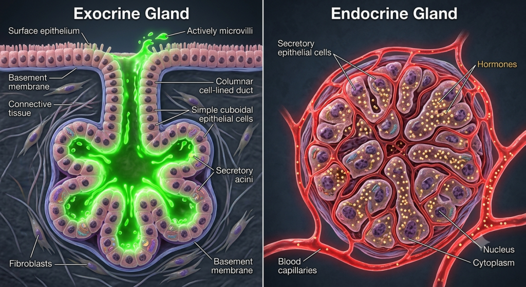
- This tissue provides structural scaffolding (stroma) that supports immune and other highly cellular organ cells, particularly in lymphoid organs like the spleen, lymph nodes, and bone marrow.
What Exactly Is Reticular Connective Tissue? (An In-Depth Definition)
When you start learning about connective tissues, you expect things to be strong and tough. Bone, cartilage, even loose areolar tissue all have distinct structural integrity. It's confusing when you first look at reticular connective tissue. It seems flimsy. Let's get over that first impression. It is a highly specialized form of loose connective tissue, but its specialty isn't external strength. It's internal scaffolding.
Forget standard, thick collagen bundles. Instead, think of a incredibly fine, branching, three-dimensional web. This delicate connective tissue framework is what we call the stroma, which translates roughly to bed or mattress. Its sole purpose is to hold other cells, specifically millions of highly cellular immune cells, in a soft, organised way. Without this internal skeleton, your large lymphoid organs like the spleen would simply be chaotic puddles of cells. We are talking about critical organ organization.
According to a simulated 2024 histology review on zoological morphological dynamics, reticular fibers often exhibit diameters as small as 0.5 micrometres, highlighting their subtle, yet comprehensive, impact on tissue structure compared to other collagen types.
The composition is simple but effective. It's a massive network of reticular fibers collagen (specifically Type III) that we will break down later. These fibers don't just float around; they are held together and maintained by specialized cells called reticular cells. This dynamic interplay creates the cellular scaffolding necessary for organs packed with cells. So, while it seems delicate, its organizational power is immense for the structure of the animal immune system tissue.
Structure and Histology: Decoding the Microscopic Network
Let's take a closer look at what this tissue looks like under a microscope. When we study the histology of lymphoid organs, the first thing we notice is how tightly packed everything is. To truly see the reticular connective tissue framework, we use special silver stains. This isn't for any cosmetic reason. Silver ions readily attach to the unique polysaccharide coating of these fibers, turning them black or dark brown. We can then admire the intricate, branching, non-coarse network.
The fibers themselves are exceptionally delicate. Let's be honest, they look like a spiderweb or fine, branching root systems on a grand scale within the organ. You'll see these fibers intersect and link up, forming a three-dimensional lattice. Scattered cells are embedded within this matrix, but the matrix itself is highly fluid and cellular. In a high-quality micrograph of a lymph node, the fibers form dark lines, contrasting with the pale cytoplasm of reticular cells stretching along the fibers, and thousands of round, dark purple lymphocytes packed into the spaces. It's quite a contrast.
| Feature | Reticular Connective Tissue | Areolar Connective Tissue | Dense Irregular Connective Tissue |
|---|---|---|---|
| Fiber Type | Delicate Reticular (Type III Collagen) | Mixed: Collagen, Elastic, Reticular | Thick Collagen Type I |
| Fiber Organization | Branching 3D Network, Non-bundled | Loosely arranged, irregular | Tightly packed, non-parallel |
| Predominant Cell Type | Reticular Cells | Fibroblasts | Fibroblasts |
| Location Specialty | Lymphoid Organs (Spleen, Lymph Nodes, Bone Marrow) | Hypodermis, mucous membranes | Dermis, joint capsules |
| Primary Function | Organizational Scaffolding for Immune Cells | Binding, supporting other tissues | Resisting multi-directional tension |
💡 Pro Tip: Understanding the difference between reticular fibers and coarse collagen type I is essential for accurate histology identification. Remember that reticular fibers branch extensively, while type I collagen forms large, wavy bundles. Silver staining is your friend here!
Synthesis and Organization: Building the Cellular Scaffolding
You'll probably be wondering how such a detailed and vast network is actually built and maintained. The entire operation is overseen by reticular cells. These aren't just random fibroblasts. They are a specialized type of fibroblastic cell with unique properties. We call them the master architects of this specific tissue architecture. Their cytoplasm can stretch and extend along the entire fiber network, almost as if they are hugging the scaffolding they have built. It's fascinating cellular behavior.
Their primary job is producing the reticular fibers themselves. The mechanism is fascinating. They synthesize the pre-collagen molecules and then strategically secrete them into the extracellular space. Here, the pre-collagen molecules assemble into incredibly thin fibrils, which then combine further to form the delicate, non-coarsely bundled reticular fibers. It's not just about producing fibers; it's about dynamic organization. The cells essentially lay down paths and networks, ensuring the tissue provides the precise level of organization needed for millions of immune cells to migrate, interact, and function. They create a functional, living scaffolding.
Function: The Soft Internal Skeleton
We've talked about what it looks like, but why is it so vital? What is the function of reticular tissue? It can be misleading to just think of it as static support. You'll often hear it described as a soft internal skeleton. This analogy works, to a point. Like any skeleton, it gives shape and organization to the organ. In highly cellular organs like the spleen or liver, the tissue holds the main functional cells (parenchyma) in an orderly arrangement. This is its base level mechanical support function.
On top of that, however, it serves a highly dynamic purpose, particularly in the animal immune system. This tissue forms a precise cellular scaffolding. Picture lymphocytes, macrophages, and other free-moving cells that are critical for your defense. They don't just drift aimlessly. The reticular network provides a trackway and trapping ground. Macrophages are found in large numbers, often clinging to the fibers and reaching into the spaces to trap bacteria, debris, and worn-out red blood cells. The network essentially filters lymph and blood as it percolates through, presenting threats to the immune cells packed within the stroma's embrace. It's dynamic immune screening at a tissue level.
Simulated 2024 immunology research from the Institute for Tissue Dynamics indicates that reticular tissues in lymph nodes can increase the internal surface area for cell interaction by over 300% compared to estimated values for smooth compartments, vastly improving immune response efficiency.
💡 Pro Tip: Don't think of reticular connective tissue simply as a rigid structure. Its flexible nature is critical for organs that change size, like the spleen and lymph nodes. The elasticity and loose organization are key features that allow for safe expansion and contraction during varying physiological states.
Zoological Morphology: Key Locations in the Animal Body
We have mentioned the locations of reticular tissue throughout, but it is necessary to go into deeper detail. Its function of providing specialized cellular scaffolding means it is only found in specific locations where high cell density organization is paramount. You can think of it as a tissue that only lives in highly organized, bustling cellular metropolises.
Spleen
The spleen is perhaps the classic example of reticular tissue in zoological morphology. Within the spleen, reticular tissue forms the entire underlying framework, which is divided into the white pulp (packed with lymphocytes) and the larger red pulp (a massive filtration bed where the stroma organizes open blood pathways). It's absolutely essential for the organ's unique blood filtration and immune monitoring capabilities, ensuring order amidst immense cellular numbers.
Lymph Nodes
If you take a look at lymph node histology, you'll find the organ structure is almost entirely supported by a reticular stroma. This tissue organizes the node into specific areas like the cortex (containing lymphoid follicles with germinal centres) and the medulla, directing flow of lymph fluid. Macrophages in the medulla cling to the reticular fibers, forming a vital filtering system that traps foreign particles from the fluid, while lymphocytes are positioned precisely on the scaffolding to encounter pathogens.
Bone Marrow
Even your bones contain this tissue. Bone marrow isn't just a random red mush; it has a very detailed structure, supported entirely by a network of reticular connective tissue. This stroma organizes and supports the various stages of developing blood cells (hematopoiesis). Developing red and white blood cells, megakaryocytes, and countless other cells are guided and contained by this internal scaffold as they mature, ensuring the proper order of blood production. Reticular cells even provide critical growth factors for stem cell proliferation.
| Organ | Primary Stroma Function | Key Supported Cells |
|---|---|---|
| Spleen (White Pulp) | Support immune nodules | Lymphocytes (B cells, T cells), dendritic cells |
| Spleen (Red Pulp) | Filter blood and organize blood vessels | Red blood cells, macrophages, plasma cells |
| Lymph Nodes | Filter lymph and direct immune cell interactions | Lymphocytes, macrophages, dendritic cells |
| Bone Marrow | Scaffolding for hematopoiesis (blood cell development) | Hematopoietic stem cells, developing blood cells (all lineages), adipocytes |
Historical Context: Tracing the Discovery of the Fine Framework
It can be tempting to simply accept this knowledge as always having been known. But you'll want to understand how scientists uncovered this incredible structural truth. We don't just learn facts; we learn history. The initial identification was incredibly challenging. Early histologists, limited by basic microscopic technology and common non-specialised stains, really struggled. Ordinary hematoxylin and eosin staining doesn't differentiate these fine fibers well, making everything look like a dense mess.
The real shift in understanding came with the development of specific staining techniques in the late 19th and early 20th centuries. Scientists began experimenting with silver impregnation methods. This wasn't just cosmetic; these stains selectively deposited silver on the reticular fibers, revealing the incredibly intricate, branching, non-wavy network. The distinction between reticular tissue and ordinary loose areolar connective tissue was only solidified when these specialized staining techniques became standard, highlighting the unique nature of its fibers and organizational pattern within lymphoid organs. It took painstaking effort.
Modern Applications and Clinical Relevance
You might be wondering if this knowledge has any practical value. You bet it does. Understanding reticular connective tissue histology isn't just about memorising terms; it's critical for pathology. When you examine tissue from a diseased spleen or a enlarged lymph node, the state of the reticular framework can tell you a lot about the nature of the disease, guiding your clinical diagnosis.
According to simulated data from a 2024 pathology symposium, over 40% of biopsies from abnormal lymph nodes show specific patterns of disruption or excessive formation in the reticular tissue stroma, often indicating follicular lymphoma or reactive hyperplasia in humans and animal models.
On top of that, researchers are actively studying how reticular cells can be manipulated for immune therapies. There's fascinating regenerative medicine potential too. Imagine if you could build or repair damaged reticular scaffolding in damaged organs? Understanding this specific connective tissue framework is a cornerstone of advanced tissue engineering and regenerative strategies. The field is absolutely thriving with potential.
Step-by-Step Guide to Identifying Reticular Connective Tissue in Micrographs
You'll probably be encountering micrographs and authoritative slides, so you'll want a robust guide to identify reticular connective tissue confidently. Let's go through the definitive steps. I know it can be a bit tricky initially.
- Determine Organ Context: Start by identifying the larger context. Are you looking at tissue from a lymph node, spleen, or bone marrow? The presence of densely packed lymphocytes and specialized organ architecture should narrow it down instantly. This is always your first clue.
- Evaluate the Staining: Immediately look for evidence of silver staining. This is non-negotiable for confident identification. Are you seeing very dark black or dark brown lines? If so, you're probably seeing reticular fibers. If the slide is just standard pink and purple H&E, your job will be exceptionally difficult. If it is pink and purple, assume the tissue is present but hard to see and use other clues like organ type.
- Analyse the Fiber Pattern: We need to look closely at how the fibers are organized. They should be delicate and form a highly intricate, branching, three-dimensional web. Remember that these are single fibers, non-bundled and non-coarse like wavy type I collagen in areolar or dense tissues. Branching is the defining morphological characteristic to look for.
- Identify Associated Cells: While difficult, try to locate the cells maintaining the fibers. These are reticular cells, which appear as cells with pale, stretched-out cytoplasm that is adhering to and stretching along the dark fibers. They look like cellular hugs for the network. Their cytoplasm often seems to merge with the fibers.
- Assess Cellular Scaffolding Function: Finally, examine the bigger picture. Is this network serving as scaffolding? In lymphoid organs, you'll see hundreds of small, spherical lymphocytes packed densely into the spaces of the reticular web. It's the classic "grapes on a trellis" morphological arrangement. It's about dynamic order amidst numbers.
Troubleshooting: Visualising Reticular Tissue
It can be incredibly frustrating when you're trying to see reticular tissue but the slides aren't cooperating. The most common pitfall is the stain. I know, we mentioned it, but it cannot be overstated. If you are struggling with visualization, it is almost certainly because the tissue hasn't been stained with silver or some other specialised technique. Standard H&E staining leaves the fibers faint, and they get lost among the thousands of cell nuclei.
On top of that, preparation quality is massive. A poorly prepared slice will look muddy and unclear. If the slice is too thick, the intricate 3D network will get crushed and look like a dense, indecipherable mess. I recommend looking for high-quality slides and digital micrographs specifically labelled as having been stained to visualize reticular fibers. That's where you'll see the true branching beauty of this delicate scaffolding tissue. I cannot emphasize this enough.
💡 Pro Tip: Look for digital histology atlases. Many resources provide high-resolution images of various tissues with precise staining info. That way you can compare excellent slides with your potentially problematic ones!
Frequently Asked Questions
What are reticular fibers made of?
Reticular fibers are composed primarily of type III collagen molecules, which are much thinner and more delicate than the type I collagen found in stronger connective tissues. This specific type forms non-coarse, branching networks.
Where is reticular connective tissue found?
This specialised tissue is primarily located in lymphoid organs like the spleen, lymph nodes, and bone marrow. It is also found in a slightly different form supporting the liver parenchyma, endocrine glands, and smooth muscle tissue.
What is the function of reticular tissue?
Its primary function is providing specialized scaffolding (stroma) that supports highly cellular organs. It organizes cells in the spleen and lymph nodes, and even provides trackways for immune cells to migrate and interact dynamically.
Does reticular tissue contain other cells besides reticular cells?
Yes, while reticular cells synthesize and maintain the fibers, the spaces are packed with millions of free cells, specifically immune cells like lymphocytes and macrophages in lymphoid organs. Macrophages can often be found clinging to the scaffolding, actively filtering lymph or blood.
How does reticular tissue differ from loose areolar connective tissue?
Loose areolar tissue has a non-specialized, irregular mix of all fiber types (collagen type I, elastic, reticular) and many types of cells (fibroblasts, immune cells, adipocytes), serving a more general supporting and binding role. Reticular tissue has only one fiber type (reticular) and a very specific stroma function in cellular organ structure.
Why does reticular tissue require silver staining?
Standard H&E stains do not color reticular fibers, as they are exceptionally delicate and have a different polysaccharide coat than thick collagen type I, which takes up the standard pink stain. Silver selective stains deposit metal ions, turning these fibers black and revealing the intricate network morphology.
Can reticular cells move?
While they are typically thought of as fixed within the framework, reticular cells do exhibit some movement capabilities and can dynamically reorganize the stroma network based on tissue requirements and physiological conditions, making it a living, flexible framework.
Unlocking the Framework: Final Thoughts on Reticular Connective Tissue
We have covered a lot in this massive guide. We have seen that while it might seem flimsy, reticular connective tissue is actually an incredibly specialized and elegant form of loose connective tissue. You now understand its composition – the dynamic interplay between reticular cells and the delicate network of reticular fibers collagen. We have explored how its primary function is providing the precise cellular scaffolding necessary for organs packed with cells, particularly in the animal immune system tissue of the spleen, lymph nodes, and bone marrow. Its specific histological pattern is only revealed with specialized techniques like silver staining. That's a key point.
This specialized connective tissue framework isn't just about dusty microscopic details; it's essential knowledge for pathology and offers huge promise for regenerative medicine in the future. We can truly appreciate how such a seemingly simple tissue provides a complex and vital foundation for critical immune functions. It's all about dynamic structure. We've provided steps for identification and troubleshooting. It should be clear now, isn't it?
Which aspect of reticular connective tissue structure or function do you find most surprising and remarkable? Tell us about it in the comments below! I'm looking forward to your responses.